. 2003 Dec 15;198(12):1863-73.
doi: 10.1084/jem.20030729. Epub 2003 Dec 8.

Mimicry of pre-B cell receptor signaling by activation of the tyrosine kinase Blk

Affiliations

Mimicry of pre-B cell receptor signaling by activation of the tyrosine kinase Blk

Theresa Tretter et al. J Exp Med. .

Abstract

During B lymphoid ontogeny, assembly of the pre-B cell receptor (BCR) is a principal developmental checkpoint at which several Src-related kinases may play redundant roles. Here the Src-related kinase Blk is shown to effect functions associated with the pre-BCR. B lymphoid expression of an active Blk mutant caused proliferation of B progenitor cells and enhanced responsiveness of these cells to interleukin 7. In mice lacking a functional pre-BCR, active Blk supported maturation beyond the pro-B cell stage, suppressed VH to DJH rearrangement, relieved selection for productive heavy chain rearrangement, and stimulated kappa rearrangement. These alterations were accompanied by tyrosine phosphorylation of immunoglobulin beta and Syk, as well as changes in gene expression consistent with developmental maturation. Thus, sustained activation of Blk induces responses normally associated with the pre-BCR.


Figures

Figure 1.
Figure 1.
(A) Overrepresentation of B220+ CD43int cells in bone marrow of Blk(Y495F) transgenic mice. Bone marrow cell suspensions from 3–4-wk-old transgenic or nontransgenic littermates were stained for B220 and additional surface markers as indicated. Plots of BP-1 or CD22 versus CD43 were gated on B220+ cells. Numbers indicate percentages of cells in the corresponding quadrants. (B) Hyperproliferation of Blk(Y495F) transgenic B cell progenitors. Bone marrow cells from transgenic or nontransgenic littermates were labeled with PKH26 and maintained in the presence of 20 ng/ml IL-7. At 3 d, cells were counterstained for B220, CD43, and BP-1. Top and middle panels show PKH-26 fluorescence gated on nontransgenic B220+ CD43+ or B220+ CD43 populations. Bottom panel shows PKH26 fluorescence gated on the transgenic B220+ CD43int population. (C) Hyperresponsiveness of Blk(Y495F) transgenic B cell progenitors to IL-7. Proliferation of B cell progenitors from nontransgenic (▴) or transgenic (▪) littermates in response to IL-7 was assayed by [3H]thymidine incorporation (mean ± SEM of three independent trials) as described in Materials and Methods.
Figure 2.
Figure 2.
Expression of Blk(Y495F) circumvents developmental blocks in RAG-deficient or μMT/μMT mice. (A) Bone marrow cell suspensions were prepared from 3–5-wk-old RAG-2−/− mice bearing the Blk(Y495F) transgene (right), or from age-matched, nontransgenic RAG-2−/− littermates (left). Cells were stained with an anti-B220 antibody and counterstained with antibodies for additional surface markers as indicated. Numbers indicate percentages of cells in the corresponding quadrants. (B) Analysis, as in A, of bone marrow cells from 3–5-wk-old μMT/μMT mice bearing Blk(Y495F) transgene (right), or from age-matched, nontransgenic μMT/μMT littermates (left).
Figure 3.
Figure 3.
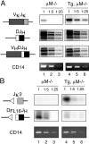
Activation of κ rearrangement and suppression of VH to DJH rearrangement in μMT/μMT mice expressing the Blk(Y495F) transgene. (A) Assay for completed rearrangements at Igμ and κ loci. VκJκ, DJH, or VHDJH rearrangements were assayed by PCR in bone marrow cells from Blk(Y495F) transgenic (lanes 4–6) or nontransgenic (lanes 1–3) μMT/μMT mice. Samples were diluted serially fivefold before amplification. Products were separated by gel electrophoresis and detected by hybridization to radiolabeled, locus-specific probes. Amplification of a nonrearranging locus (CD14, bottom) was performed as a control. (B) Assay for DNA cleavage at V(D)J recombination signal sequences. Double strand DNA breaks at the Jκ2 recombination signal sequence (top) or the 5′ recombination signal sequence of DFL16.1 (middle) were detected in bone marrow DNA from nontransgenic (lanes 1–3) or transgenic (lanes 4–6) μMT/μMT mice by ligation-mediated PCR. Samples were diluted serially fivefold before amplification. Products were detected as in A.
Figure 4.
Figure 4.
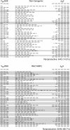
Blk(Y495F) relieves selection for productive heavy chain gene rearrangement. Nucleotide sequences of VHDJH junctions from nontransgenic or Blk(Y495F) transgenic littermates are shown. Genomic DNA was purified from sorted B220+ CD43 bone marrow cells of 3-wk-old nontransgenic or Blk(Y495F) transgenic mice. VHJ558-D-JH3 junctions were amplified by PCR. Nucleotide sequences of individual, cloned junctions are displayed. The 3′ and 5′ ends of germline VHJ558 and JH3 segments, respectively, are shown at the top. For each entry below, the 3′ end of sequence derived from VHJ558 and the 5′ end of sequence derived from JH3 are separated by sequence derived from the D segment and any N or P nucleotide additions. Nonproductive rearrangements are shaded. The difference in abundance of nonproductively rearranged alleles from transgenic and nontransgenic mice was highly significant (P < 0.000001).
Figure 5.
Figure 5.
Appearance of cells bearing a B progenitor phenotype in spleens of Blk(Y495F) transgenic mice. (A) Flow cytometric analysis. Single cell suspensions from spleens of transgenic (left) or nontransgenic (right) littermates (3–5 wk old) were stained with antibodies to the indicated markers. Plots of BP-1, CD22, or CD43 versus IgM (bottom three pairs) are gated on B220+ cells. Numbers indicate the percentage of cells in the corresponding quadrant. (B) Peripheral expression of immature B cell markers in Blk(Y495F) transgenic mice. RNA was prepared from spleens of nontransgenic (lanes 1–3) or transgenic (lanes 4–6) littermates and transcripts encoding RAG-2, terminal nucleotidyl transferase (TdT), VpreB, or actin were detected by RT-PCR. Products were diluted as indicated above, fractionated by gel electrophoresis, and detected by staining with ethidium bromide.
Figure 6.
Figure 6.
Targets of proximal and distal signaling in B cell progenitors from Blk(Y495F) transgenic mice. (A) Constitutive tyrosine phosphorylation of Igβ in transgenic B cell progenitors. Lysates were prepared from pro–B cells of nontransgenic (lanes 1, 4, and 7) or transgenic (lanes 2, 5, and 8) mice, and Igβ was immunoprecipitated (lanes 4–8). Control immunoprecipitations were performed from a thymocyte lysate (lanes 3 and 6) or with nonimmune IgG (lanes 1–3). Undiluted (lanes 1–6) and twofold diluted (lanes 7 and 8) immunoprecipitates were fractionated by electrophoresis alongside whole cell lysates (lanes 9–11). Phosphotyrosine (top) and Ig-β (bottom) were detected by sequential immunoblotting. Arrows mark the position of Igβ. (B) Constitutive tyrosine phosphorylation of Syk in transgenic B cell progenitors. Lysates were prepared from nontransgenic (lanes 1 and 2) or transgenic (lanes 3 and 4) pro–B cells as in A. Syk was immunoprecipitated (lanes 2 and 4) and control immunoprecipitations were performed with nonimmune IgG (lanes 1 and 3). Phosphotyrosine (top) and Syk (bottom) were detected by sequential immunoblotting. (C) Differential gene expression in pro–B cells from transgenic and nontransgenic mice. 35 genes of known function that were differentially expressed in nontransgenic (left) and transgenic (right) pro–B cells are shown. Each column corresponds to one microarray. Red represents expression above and green represents expression below the median value. Black represents expression at the median and gray represents no detectable expression. (D) Confirmation of differential expression. Total RNA from nontransgenic (lanes 1–4) or transgenic (lanes 5–8) pro–B cells was reverse transcribed, diluted serially fourfold, and used as a template for amplification of the transcripts indicated at right. Products were fractionated by gel electrophoresis and detected with ethidium bromide.

References

    1. Gellert, M. 2002. V(D)J recombination: RAG proteins, repair factors and regulation. Annu. Rev. Biochem. 71:101–132. - PubMed
    1. Melchers, F., E. ten Boekel, T. Seidl, X.C. Kong, T. Yamagami, K. Onishi, T. Shimizu, A.G. Rolink, and J. Andersson. 2000. Repertoire selection by pre-B-cell receptors and B-cell receptors, and genetic control of B-cell development from immature to mature B cells. Immunol. Rev. 175:33–46. - PubMed
    1. Reth, M., and J. Wienands. 1997. Initiation and processing of signals from the B cell antigen receptor. Annu. Rev. Immunol. 15:453–479. - PubMed
    1. Stoddart, A., H.E. Fleming, and C.J. Paige. 2000. The role of the preBCR, the interleukin-7 receptor, and homotypic interactions during B-cell development. Immunol. Rev. 175:47–58. - PubMed
    1. Fleming, H.E., and C.J. Paige. 2001. Pre-B cell receptor signaling mediates selective response to IL-7 at the pro-B to pre-B cell transition via an ERK/MAP kinase-dependent pathway. Immunity. 15:521–531. - PubMed

Publication types