. 2003 Dec 15;198(12):1797-806.
doi: 10.1084/jem.20030735. Epub 2003 Dec 8.

Interleukin 7 regulates the survival and generation of memory CD4 cells

Affiliations

Interleukin 7 regulates the survival and generation of memory CD4 cells

Robyn M Kondrack et al. J Exp Med. .

Abstract

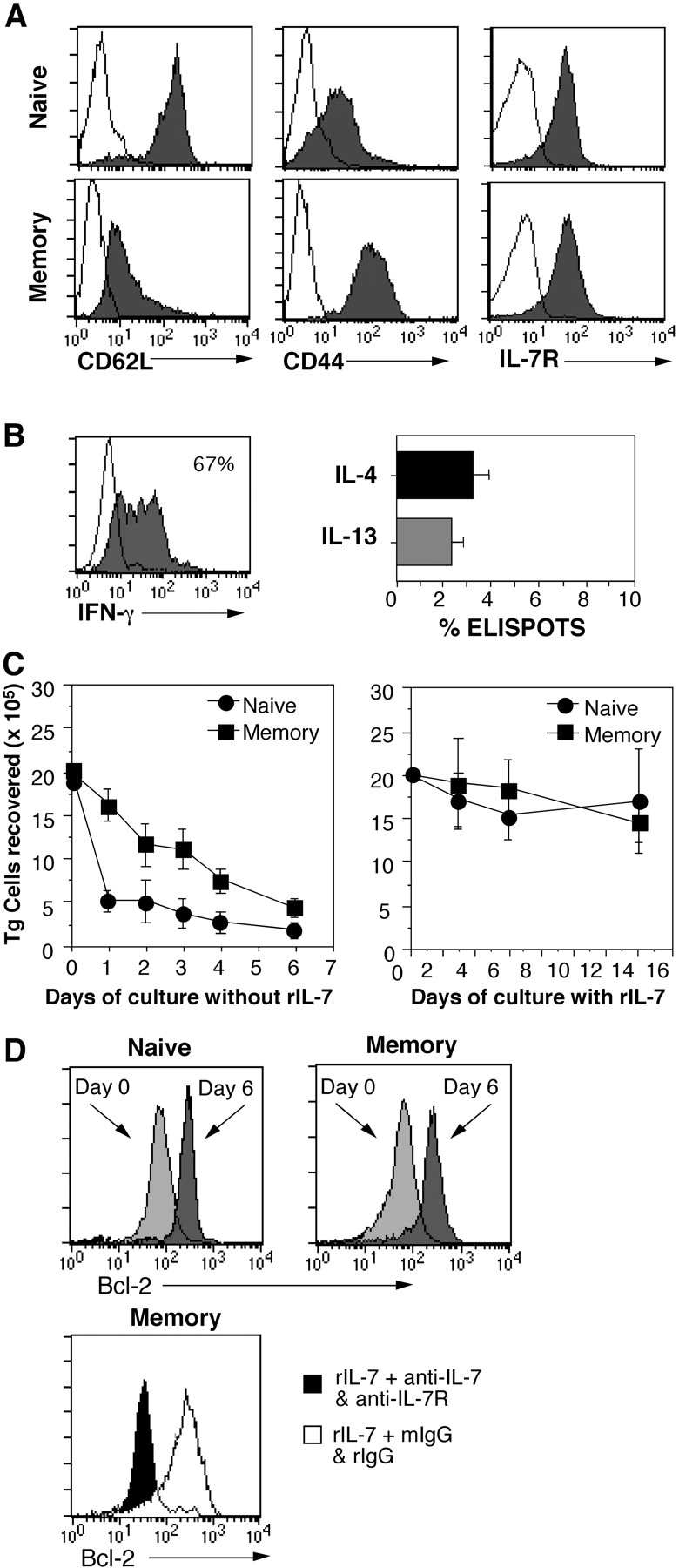
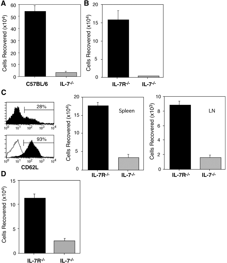
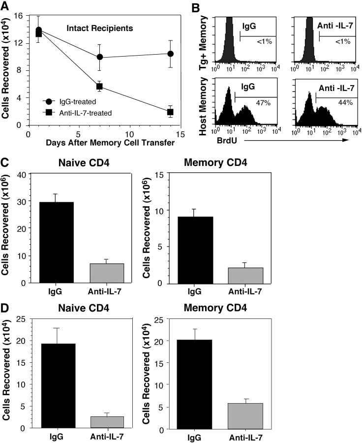
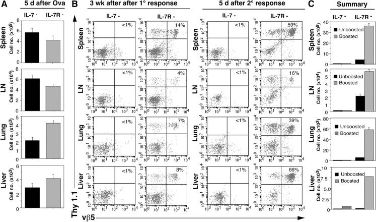
Cytokines, particularly those of the common gamma chain receptor family, provide extrinsic signals that regulate naive CD4 cell survival. Whether these cytokines are required for the maintenance of memory CD4 cells has not been rigorously assessed. In this paper, we examined the contribution of interleukin (IL) 7, a constitutively produced common gamma chain receptor cytokine, to the survival of resting T cell receptor transgenic memory CD4 cells that were generated in vivo. IL-7 mediated the survival and up-regulation of Bcl-2 by resting memory CD4 cells in vitro in the absence of proliferation. Memory CD4 cells persisted for extended periods upon adoptive transfer into intact or lymphopenic recipients, but not in IL-7- mice or in recipients that were rendered deficient in IL-7 by antibody blocking. Both central (CD62L+) and effector (CD62L-) memory phenotype CD4 cells required IL-7 for survival and, in vivo, memory cells were comparable to naive CD4 cells in this regard. Although the generation of primary effector cells from naive CD4 cells and their dissemination to nonlymphoid tissues were not affected by IL-7 deficiency, memory cells failed to subsequently develop in either the lymphoid or nonlymphoid compartments. The results demonstrate that IL-7 can have previously unrecognized roles in the maintenance of memory in the CD4 cell population and in the survival of CD4 cells with a capacity to become memory cells.


Figures

Figure 1.
Figure 1.
In vivo–primed TCR transgenic memory CD4 cells survive in response to rIL-7 in vitro. Purified naive OT-II Thy 1.1 CD4 cells were transferred into C57BL/6 Rag2 mice (5 × 10⁶ cells/recipient) and immunized with OVA protein and adjuvant as indicated in Materials and Methods. 1 mo later, resting memory OT-II cells were isolated and compared with freshly isolated naive OT-II cells. (A) Phenotype of memory OT-II cells. Naive and memory OT-II cells were stained for expression of CD62L, CD44, and IL-7Rα and analyzed by flow cytometry (shaded histograms; unshaded histograms denote background staining). (B) Frequencies of effector cytokine producers among memory OT-II cells. Memory OT-II Thy 1.1 cells were restimulated with OVA peptide in the presence of splenic APC and tested for secretion of IFN-γ at 12 h by ICS, and after enrichment of Thy 1.1 cells, for production of IL-4 or IL-13 at 24 h by ELISPOT analysis. (C) IL-7 promotes survival of OT-II cells. Naive and memory OT-II Thy 1.1 cells were cultured at 106/ml for the indicated number of days without or with rIL-7 at 10 ng/ml (left and right, respectively). (D) Blocking IL-7 prevents up-regulation of Bcl-2. Naive (top left) and memory (top right) OT-II Thy 1.1 CD4 cells were stained for expression of Bcl-2 (light gray histograms). The cells were cultured as in C for 6 d with 10 ng/ml rIL-7 and stained for BcL-2 (dark gray histograms). Memory cell cultures were also treated with either 10 μg/ml each anti–IL-7 and anti–IL-7R mAb or with an equivalent amount of rat and mouse IgG (bottom, rIgG and mIgG, respectively). The cells were stained for Bcl-2 on day 6 after culture with blocking mAb (shaded histogram) or with control mAb (unshaded histogram).
Figure 2.
Figure 2.
Survival of memory CD4 cells in IL-7–deficient recipients. (A) Comparison with intact recipients. Resting OT-II Thy 1.1 memory CD4 cells were generated and isolated as for Fig. 1 and sorted by flow cytometry to obtain CD44hi cells. These cells were transferred into normal C57BL/6 or IL-7 mice (2 × 10⁶ cells/recipient). 1 wk later, donor cell recoveries were quantitated in the host spleens and LNs from the total nucleated cell counts and fractions of transgenic donor (vα2, vβ5, and Thy 1.1+) CD4 cells. (B) Comparison with IL-7R recipients. OT-II Thy 1.1 memory CD4 cells were isolated and transferred as in A into IL-7R and IL-7 mice, and analyzed for transgenic donor cell recovery after 1 wk. (C) Susceptibility of CD62L+ memory cells to IL-7 deprivation. OT-II Thy 1.1 memory cells were isolated from the LNs of OVA-primed Rag2 that were primed as for Fig. 1. The cells were stained for CD62L before (top) and after magnetic selection for positively expressing cells (bottom, overlay of sorted CD62L and CD62L+ subsets). CD62L+ cells were transferred into either IL-7R or IL-7 hosts (2 × 10⁶ cells/recipient), and the recovery of donor memory CD4 cells was assessed after 10 d. (D) Naive OT-II Thy 1.1 CD4 cells were primed by transfer into normal C57BL/6 mice (5 × 10⁶ each) and immunization of the recipients with OVA and adjuvant. 4 wk later, donor memory CD4 cells isolated from the spleens were transferred into IL-7R and IL-7 mice (0.5 × 10⁶ cells/recipient). These mice were evaluated 1 wk after cell transfer for the presence of transgenic donor cells in the LNs and spleens.
Figure 3.
Figure 3.
Effects blocking IL-7 on transgenic and polyclonal memory CD4 cells. (A) Blocking of transgenic memory cell survival in normal recipients. OT-II Thy 1.1 memory cells were primed in vivo and isolated as for Fig. 1. The cells were transferred into normal C57 BL/6 mice (2 × 10⁶ cells/recipient). On the day of cell transfer, separate groups of recipients were injected with 1 mg of either anti–IL-7 or mIgG. Additional doses were given every other day through day 12. On days 1, 7, and 14, animals from each group were evaluated for transgenic donor cell recovery from the spleens and LNs. (B) Lack of division of donor transgenic memory cells but expansion of host polyclonal memory cells in anti–IL-7–treated recipients. Recipients were administered BrdU as described in Materials and Methods to assess proliferation of OT-II Thy 1.1 memory cells and in a separate experiment, division of host memory phenotype (CD44hi) CD4 cells. BrdU uptake was assessed by ICS of splenic lymphocytes. Histograms gated on donor Thy 1.1, vα2, and vβ5+ cells (top) and on host CD44hi and CD4 cells (bottom) are shown. (C) Diminished recovery of host naive and memory CD4 cells. The effects of anti–IL-7 treatment on the recovery of naive phenotype (CD44lo) and memory phenotype (CD44hi) CD4 cells were assessed by quantitating Thy 1.2+ host CD4 cells from the spleens and LNs of the recipients from A on day 14. Comparable results were obtained using high versus low expression of α4 integrin to distinguish memory versus naive phenotype host CD4 cells (not depicted). (D) Recovery of naive and resting memory CD4 cells after IL-7 treatment of immunodeficient mice. Naive CD4 cells were isolated from (AND B10.BR × B6PL.Thy)F1 mice and were transferred into (B10.BR × C57BL/6) F1 SCID mice (5 × 10⁶ cells/recipient). One set of recipients was treated with anti–IL-7 or mIgG as for A without immunization. A second set of recipients was primed with PCC peptide as described in Materials and Methods. 1 mo later, when AND CD4 cells from the spleen were uniformly CD62L and CD44hi, recipients were treated either with anti–IL-7 or with mIgG as for A. Recovery of transgenic donor (vα11, vβ3, and Thy 1.1+) from the spleens and LNs was determined on day 14 after treatment for naive and primed recipients (left and right, respectively).
Figure 4.
Figure 4.
Depletion of primed CD4 cells in the absence of IL-7. (A) Cell recovery from lymphoid and nonlymphoid tissues after primary immunization. Naive OT-II Thy CD4 cells were transferred into IL-7 or IL-7R mice (5 × 10⁶ cells/recipient). The mice were immunized with OVA as described in Materials and Methods. On day 5, donor cell recovery was assessed in the spleen, LNs, lung, and liver as depicted in Fig. 2. (B) Recovery of primed CD4 cells after rest and challenge of IL-7 and IL-7R recipients. Mice from the same experiment shown in A were evaluated for the presence of donor CD4 cells at 3 wk after OT-II Thy 1.1 CD4 cell transfer and immunization (left), and 5 d later after a secondary response was induced by challenge with OVA (right). Thy 1.1 and vβ5 staining of the total CD4 cells recovered from each tissue is shown. (C) Summary. Total donor OT-II cell recovery from each site before and after boosting with OVA, as determined from the cell counts as for Fig. 2.

References

    1. Welsh, R.M., and L.K. Selin. 2002. No one is naive: the significance of heterologous T-cell immunity. Nat. Rev. Immunol. 2:417–426. - PubMed
    1. Homann, D., L. Teyton, and M.B.A. Oldstone. 2001. Differential regulation of antiviral T-cell immunity results in stable CD8+ but declining CD4+ T cell memory. Nat. Med. 7:913–919. - PubMed
    1. Varga, S.M., L.K. Selin, and R.M. Welsh. 2001. Independent regulation of lymphocytic choriomeningitis virus-specific T cell memory pools: relative stability of CD4 memory under conditions of CD8 memory T cell loss. J. Immunol. 166:1554–1561. - PubMed
    1. Ernst, B., D.-S. Lee, J.M. Chang, J. Sprent, and C.D. Surh. 1999. The peptide ligands mediating positive selection in the thymus control T cell survival and homeostatic proliferation in the periphery. Immunity. 11:173–181. - PubMed
    1. Tanchot, C.F., A. Lemonnier, B. Perarnau, and A.A. Freitas. 1997. Differential requirements for survival and proliferation of CD8 naive or memory T cells. Science. 276:2057–2062. - PubMed

Publication types