. 2003 Dec 15;198(12):1853-62.
doi: 10.1084/jem.20031223. Epub 2003 Dec 8.

A critical role for induced IgM in the protection against West Nile virus infection

Affiliations

A critical role for induced IgM in the protection against West Nile virus infection

Michael S Diamond et al. J Exp Med. .

Abstract

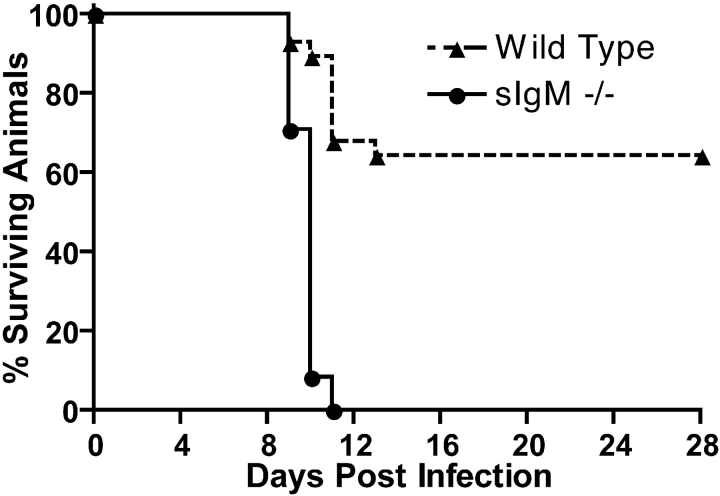
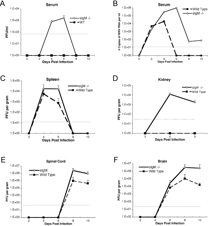
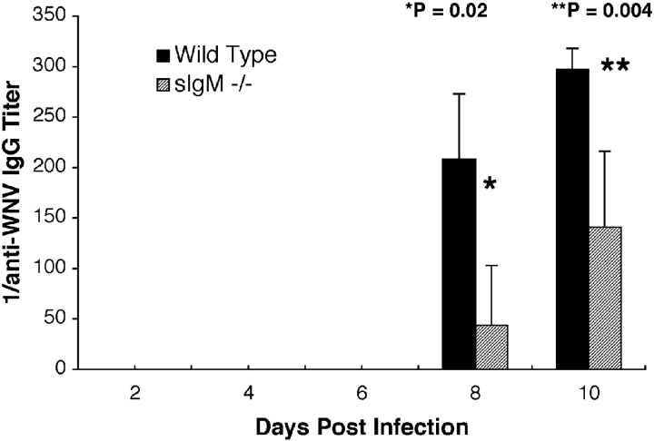
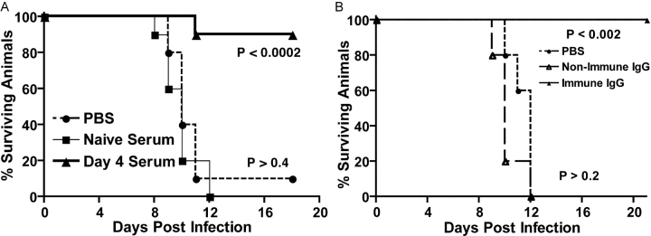
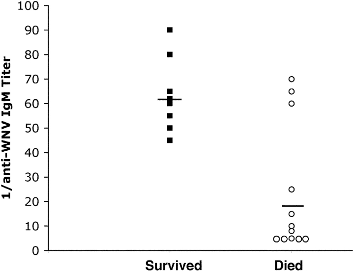
In humans, the elderly and immunocompromised are at greatest risk for disseminated West Nile virus (WNV) infection, yet the immunologic basis for this remains unclear. We demonstrated previously that B cells and IgG contributed to the defense against disseminated WNV infection (Diamond, M.S., B. Shrestha, A. Marri, D. Mahan, and M. Engle. 2003. J. Virol. 77:2578-2586). In this paper, we addressed the function of IgM in controlling WNV infection. C57BL/6J mice (sIgM-/-) that were deficient in the production of secreted IgM but capable of expressing surface IgM and secreting other immunoglobulin isotypes were vulnerable to lethal infection, even after inoculation with low doses of WNV. Within 96 h, markedly higher levels of infectious virus were detected in the serum of sIgM-/- mice compared with wild-type mice. The enhanced viremia correlated with higher WNV burdens in the central nervous system, and was also associated with a blunted anti-WNV IgG response. Passive transfer of polyclonal anti-WNV IgM or IgG protected sIgM-/- mice against mortality, although administration of comparable amounts of a nonneutralizing monoclonal anti-WNV IgM provided no protection. In a prospective analysis, a low titer of anti-WNV IgM antibodies at day 4 uniformly predicted mortality in wild-type mice. Thus, the induction of a specific, neutralizing IgM response early in the course of WNV infection limits viremia and dissemination into the central nervous system, and protects against lethal infection.


Figures

Figure 1.
Figure 1.
Survival data for C57BL/6J mice inoculated with WNV. Wild-type and sIgM−/− mice were inoculated via footpad with the indicated doses of WNV and followed for 28 d. The survival curves were constructed using data from three to five separate experiments. The number of animals were n = 28 for wild-type mice and n = 24 for sIgM−/− mice. The mean survival times (in days) were 21.8 ± 1.6 for wild-type and 9.8 ± 0.1 for sIgM−/− mice (P < 0.0001).
Figure 2.
Figure 2.
WNV burden in peripheral and CNS tissues of adult wild-type and sIgM−/− C57BL/6J mice. (A) Infectious virus levels in serum. Serum was harvested at the indicated days after subcutaneous footpad inoculation with 102 PFU of WNV and virus levels were measured using a viral plaque assay in BHK21 cells. Data are shown as the mean of PFU per milliliter of serum and reflect from 6 to 10 mice per time point. The dotted line represents the limit of sensitivity of the assay. (B) WNV RNA levels in serum. Viral RNA levels were determined from serum of wild-type or sIgM−/− mice after WNV infection at the indicated days using a real-time fluorogenic RT-PCR assay. Data are expressed as genomic equivalents of WNV RNA per milliliter of serum and reflect the mean of five independent mice per time point. (C) Infectious virus levels in the spleen. Virus levels were determined from spleen homogenates by plaque assay and normalized for the weight of the tissue sample. Data are the mean of 10 animals per time point. (D) Infectious virus levels in the kidney. Virus levels were determined from kidney homogenates by plaque assay and normalized for the weight of the tissue sample. Data are the mean of 10 kidneys per time point. (E and F) Infectious virus levels in the CNS. Virus levels were determined from spinal cord (E) or brain (F) as described in Fig. 2 C.
Figure 3.
Figure 3.
Development of specific IgG against WNV. Serum was collected from wild-type or sIgM−/− mice at the indicated days after infection. The levels of specific IgG were determined by incubating serum with adsorbed control or purified WNV E protein. Data are the mean of serum from 8 to 12 mice per time point performed in duplicate. p-values and statistical significance are as indicated.
Figure 4.
Figure 4.
Passive administration of serum or purified antibodies to sIgM−/− mice. (A) Serum was collected from naive mice, or mice that were infected with WNV for 4 d. After heat-inactivation, 0.5 ml of serum was administered to sIgM−/− mice 1 d before and after infection with 102 PFU of WNV. Data reflect two independent experiments with five mice per condition, and p-values are indicated next to each curve. (B) 10 mg of purified human immune or nonimmune IgG were administered as a single dose via an intraperitoneal route immediately before administration of 102 PFU of WNV via footpad inoculation. Data are from two independent experiments with five mice per condition, and p-values are indicated next to each curve.
Figure 5.
Figure 5.
Scatter plot of the relationship between specific anti-WNV IgM titer at day 4 after infection and survival. 7-wk-old wild-type mice were infected with 102 PFU of WNV. At day 4 after infection, animals were phlebotomized and followed clinically for survival. An ELISA was used to evaluate serum for specific IgM titer against purified WNV E protein using a biotin-conjugated goat anti–mouse IgM secondary antibody. 20 animals were included in the study. 8 survived and 12 died after infection. Five of the animals that died had identical titers of 1:5. Solid lines indicate the mean titer.

References

    1. Hubalek, Z., and J. Halouzka. 1999. West Nile fever—a reemerging mosquito-borne viral disease in Europe. Emerg. Infect. Dis. 5:643–650. - PMC - PubMed
    1. Lanciotti, R.S., J.T. Roehrig, V. Deubel, J. Smith, M. Parker, K. Steele, B. Crise, K.E. Volpe, M.B. Crabtree, J.H. Scherret, et al. 1999. Origin of the West Nile virus responsible for an outbreak of encephalitis in the northeastern United States. Science. 286:2333–2337. - PubMed
    1. Camenga, D.L., N. Nathanson, and G.A. Cole. 1974. Cyclophosphamide-potentiated West Nile viral encephalitis: relative influence of cellular and humoral factors. J. Infect. Dis. 130:634–641. - PubMed
    1. Asnis, D.S., R. Conetta, A.A. Teixeira, G. Waldman, and B.A. Sampson. 2000. The West Nile Virus outbreak of 1999 in New York: the Flushing Hospital experience. Clin. Infect. Dis. 30:413–418. - PubMed
    1. Tsai, T.F., F. Popovici, C. Cernescu, G.L. Campbell, and N.I. Nedelcu. 1998. West Nile encephalitis epidemic in southeastern Romania. Lancet. 352:767–771. - PubMed

Publication types