. 2003 Dec 23;100(26):16012-7.
doi: 10.1073/pnas.2534856100. Epub 2003 Dec 8.

Fundamental role of the Rip2/caspase-1 pathway in hypoxia and ischemia-induced neuronal cell death

Affiliations

Fundamental role of the Rip2/caspase-1 pathway in hypoxia and ischemia-induced neuronal cell death

Wen-Hua Zhang et al. Proc Natl Acad Sci U S A. .

Erratum in

  • Proc Natl Acad Sci U S A. 2004 Feb 3;101(5):1426

Abstract

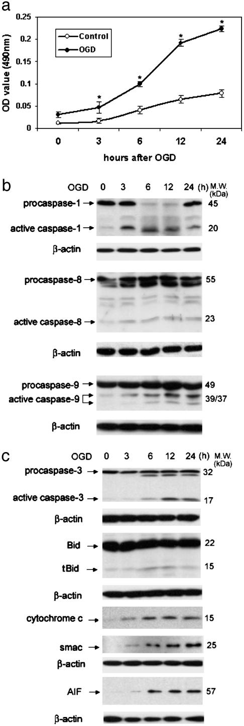
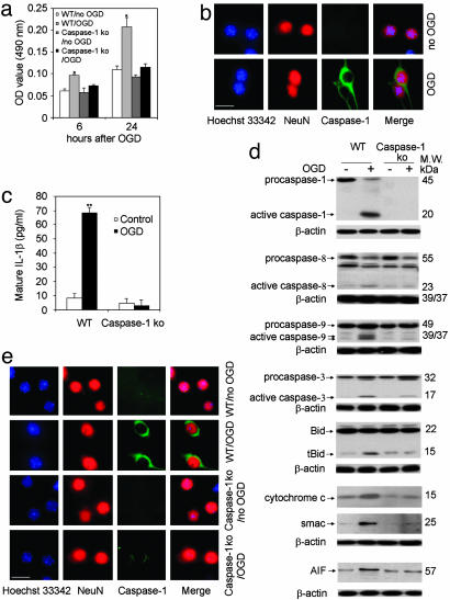
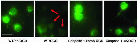
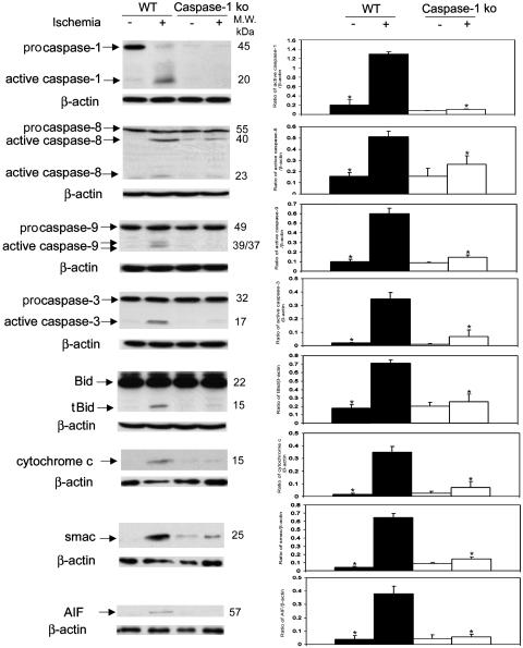
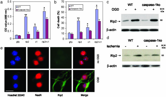
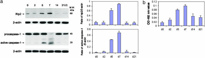
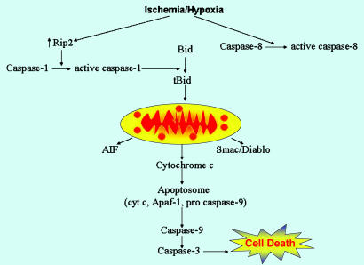
Caspase-1 plays a key role in inflammatory pathways by processing pro-IL-1beta into the active cytokine mature IL-1beta. Given its sequence similarity with the Caenorhabditis elegans cell death gene ced-3,it has long been speculated that caspase-1 may also play a role in cell death. However, an unequivocal role for caspase-1 in cell death has been questioned, and not definitively demonstrated. Furthermore, if caspase-1 does play a role in cell death, its position in the apoptotic hierarchy has not been clearly defined. Previous studies have shown that caspase-1 knockout (KO) mice and transgenic mice expressing a dominant-negative caspase-1 construct are resistant to ischemic brain injury. We provide direct evidence that caspase-1 plays a key role in neuronal cell death and that caspase-1 is an apical activator of the cell death pathway in the premitochondrial collapse stage. Furthermore, we demonstrate that Rip2/Cardiak/Rick is a stress-inducible upstream modulator of pro-caspase-1 apoptotic activation. We provide evidence that Bid cleavage appears to be an important downstream effector of caspase-1-mediated cell death. Our data demonstrate that caspase-1 is an apical mediator of neuronal cell death during in vitro hypoxia, and confirmed in vivo in ischemia, and provide insights into the sequence of events involved in this pathological cell death process.


Figures

Fig. 1.
Fig. 1.
Activation of caspase-1, -3, -8, and -9, cleavage of Bid, and release of cytochrome c, Smac/Diablo, and AIF during OGD-induced neuronal cell death. (a) Primary cortical neurons from wild-type mice cultured for a week were treated with 3 h of OGD. At the indicated time points after completion of OGD, culture media was collected and analyzed for LDH release as an indicator of cell death. Results are the average of three independent experiments. *, P < 0.05 versus control. (b) Cell extracts were subjected to immunoblotting with antibodies against caspase-1, -3, -8, and -9 and Bid. Cytosolic extracts were evaluated for release of cytochrome c, Smac/Diablo, AIF, and β-actin. The blots are representative of three independent experiments.
Fig. 2.
Fig. 2.
Role of caspase-1 in OGD-induced neuronal death. (a) Primary cortical neurons from wild-type mice and caspase-1 KO mice cultured for a week were treated with 3 h of OGD. Supernatants were collected and assayed for LDH release 6 and 24 h after the completion of OGD. Results are the average of three independent experiments. *, P < 0.05 versus control. (b) Six hours after completion of OGD, wild-type cells were processed for immunofluorescence stained for active caspase-1 (green). Blue, nuclei stained by Hoechst; red, neurons stained with NeuN. (Scale bar, 2.5 μm.) (c-e) Wild-type and caspase-1 KO neurons were treated with OGD. (c) Supernatants were collected and assayed for mature IL-1β release 6 h after the completion of OGD. Results are the average of three independent experiments. **, P < 0.01 versus control. (d) Cell extracts were subjected to immunoblotting with antibodies against caspase-1, -3, -8, and -9, bid, cytochrome c, Smac/Diablo, AIF, and β-actin. The blots are representative of three independent experiments. (e) Cells were also processed for immunofluorescence staining with antibodies against active caspase-3 (green). Blue, nuclei stained by Hoechst; red, neurons stained with NeuN. (Scale bar, 2.5 μm.)
Fig. 3.
Fig. 3.
Caspase-1-deficient neurons demonstrate resistance to hypoxia-mediated loss of mitochondrial membrane potential. Live primary cortical neurons were stained directly with 2 μM Rhodamine-123 over 6 h after 3 h of OGD. Arrows show loss of mitochondrial transmembrane potential after OGD treatment of wild-type cortical neurons. Mitochondrial transmembrane potential is retained after OGD in caspase-1-deficient neurons. (Scale bar, 2.5 μm.)
Fig. 4.
Fig. 4.
Role of caspase-1 in ischemia-induced neuronal cell death. Caspase-1 KO mice and wild-type mice were subjected to 2 h of focal ischemia. After 5 h of reperfusion, the ischemic territory was isolated. Whole tissue lysates were subjected to immunoblotting with antibodies against caspase-1, -3, -8, and -9 and Bid. Cytosolic extracts of ischemic tissue were evaluated for release of cytochrome c, Smac/Diablo, and AIF. (Right) Densitometry with three samples for each condition. *, P < 0.05 versus wild-type ischemia group.
Fig. 5.
Fig. 5.
Role of Rip2 in caspase-1-mediated neuronal cell death. Primary cortical neurons were transfected with caspase-1 (c1), Rip2, or both. Forty-eight hours after transfection, media were collected and assayed for LDH release (a) and evaluation of cell death (b). Results are the average of three independent experiments. *, P < 0.05 versus caspase-1 KO. (c and d) The same samples as in Figs. 2d and 4 were subjected to immunoblotting with antibodies against Rip2 and β-actin. (e) The same cells as Fig. 2b used were also processed for immunofluorescence staining with antibodies against Rip2 (green). Blue, nuclei stained by Hoechst; red, neurons stained with NeuN. (Scale bar, 2.5 μm.)
Fig. 6.
Fig. 6.
Rip2 up-regulation results in caspase-1 activation. Primary cortical neurons from wild-type mice cultured for 0, 2, 5, 7, 14, and 21 days were treated with 3 h OGD. (a) Cells were collected 6 h after OGD and lysates were immunoblotted with antibodies against Rip2, caspase-1, and β-actin. (Right) Densitometry using three samples for each condition. (b) Supernatants were assayed for LDH release.
Fig. 7.
Fig. 7.
The Rip2/Caspase-1 pathway plays a key role in ischemia/hypoxia-associated neuronal death. From the above-described data, a pathway mediating neuronal death involving Rip2 and caspase-1 may be described. After the hypoxia/ischemia insult, increased levels of Rip2 protein are detected within neurons. Rip2 then mediates caspase-1 activation. Caspase-1 thereafter generates the proapoptotic tBid fragment. tBid plays a role in the release of mitochondrial apoptogenic factors, including cytochrome c, AIF, and Smac/Diablo. Released cytochrome c induces apoptosome assembly, resulting in caspase-9 and -3 activation. Activated caspase-3 plays a key role in the final stages of the apoptotic cascade. In our in vivo and in vitro models, we do detect caspase-8 activation. However, it appears that the magnitude of caspase-8 activation is not sufficient to be of functional importance. Furthermore, the mechanism resulting in ischemia/hypoxia-mediated caspase-8 activation, whether it is direct or indirect, is not clearly understood.

References

    1. Thornberry, N. A., Rano, T. A., Peterson, E. P., Rasper, D. M., Timkey, T., Garcia-Calvo, M., Houtzager, V. M., Nordstrom, P. A., Roy, S., Vaillancourt, J. P., et al. (1997) J. Biol. Chem. 272, 17907-17911. - PubMed
    1. Shi, Y. (2002) Mol. Cell 9, 459-470. - PubMed
    1. Wang, S., Miura, M., Jung, Y. K., Zhu, H., Li, E. & Yuan, J. (1998) Cell 92, 501-509. - PubMed
    1. Nakagawa, T., Zhu, H., Morishima, N., Li, E., Xu, J., Yankner, B. A. & Yuan, J. (2000) Nature 403, 98-103. - PubMed
    1. Li, M., Ona, V. O., Guegan, C., Chen, M., Jackson-Lewis, V., Andrews, L. J., Olszewski, A. J., Stieg, P. E., Lee, J. P., Przedborski, S. & Friedlander, R. M. (2000) Science 288, 335-339. - PubMed

Publication types

MeSH terms

Substances