. 2003 Dec 23;100(26):15583-8.
doi: 10.1073/pnas.2531786100. Epub 2003 Dec 8.

Membrane tube formation from giant vesicles by dynamic association of motor proteins

Affiliations

Membrane tube formation from giant vesicles by dynamic association of motor proteins

Gerbrand Koster et al. Proc Natl Acad Sci U S A. .

Abstract

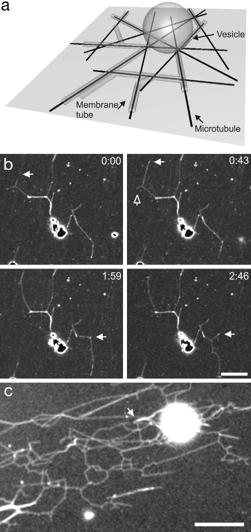
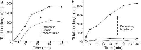
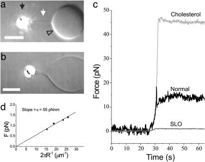
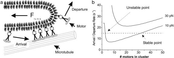
The tubular morphology of intracellular membranous compartments is actively maintained through interactions with motor proteins and the cytoskeleton. Moving along cytoskeletal elements, motor proteins exert forces on the membranes to which they are attached, resulting in the formation of membrane tubes and tubular networks. To study the formation of membrane tubes by motor proteins, we developed an in vitro assay consisting of purified kinesin proteins directly linked to the lipids of giant unilamellar vesicles. When the vesicles are brought into contact with a network of immobilized microtubules, membrane tubes and tubular networks are formed. Through systematic variation of the kinesin concentration and membrane composition we study the mechanism involved. We show that a threshold concentration of motor proteins is needed and that a low membrane tension facilitates tube formation. Forces involved in tube formation were measured directly with optical tweezers and are shown to depend only on the tension and bending rigidity of the membrane. The forces were found to be higher than can be generated by individual motor proteins, indicating that multiple motors were working together to pull tubes. We propose a simple mechanism by which individual motor proteins can dynamically associate into clusters that provide the force needed for the formation of tubes, explaining why, in contrast to earlier findings [Roux, A., Cappello, G., Cartaud, J., Prost, J., Goud, B. & Bassereau, P. (2002) Proc. Natl. Acad. Sci. USA 99, 5394-5399], motor proteins do not need to be physically linked to each other to be able to pull tubes.


Figures

Fig. 1.
Fig. 1.
Overview of the experimental system. (a) Schematic representation of the assay (not to scale). Membrane tubes are formed from a vesicle that lies on top of a random network of MTs attached to the surface. (b) Time sequence of scanning confocal microscopy images of membrane tubes during the early stage of network formation (≈10 min after sample preparation). The network is dynamic: existing tubes disappear (open arrow) and new tubes appear and grow (white arrows), giving shape to three-way junctions. The fluorescence is due to fluorescently labeled streptavidin. Neither MTs nor motor proteins are visible. Time is given in minutes and seconds. (Bar, 5 μm.) (c) Fluorescence image of a large network of membrane tubes (with streptolysin). After 2 h, multiple three-way junctions can be observed and multiple membrane tubes are formed alongside each other, as can be seen from a stepwise increase in fluorescence (see arrow). Membranes are stained with BODIPY. (Bar, 10 μm.)
Fig. 2.
Fig. 2.
Evolution of tubular networks. (a) A typical example of the evolution of the total length of membrane tubes pulled from vesicles for different final concentrations of kinesin in the sample: 10 μg/ml (filled circles), 3 μg/ml (open squares), 1 μg/ml (open circles), and 0.1 μg/ml (filled squares). In each sample, ≈15 vesicles were present in the field of view. Time 0 corresponds to the end of sample preparation. No tubes are formed at the lower concentrations, whereas, for the higher concentrations, tubes start to form after sedimentation. (b) Typical example of the total tube length pulled from vesicles with the pore-forming drug SLO and cholesterol (filled circles), normal vesicles (filled squares), and vesicles with only cholesterol (open circles). In the presence of SLO the forces needed to pull tubes are much lower than for normal vesicles, whereas with only cholesterol present they are higher (see Fig. 3). The kinesin concentration is 2 μg/ml.
Fig. 3.
Fig. 3.
Force measurement of tube formation. (a) Video-enhanced differential interference contrast image of a normal vesicle (open arrow) from which a tube (white arrow) is pulled with a bead (black arrow) held in optical tweezers. The contrast has been enhanced to make the tube visible. (Bar, 5 μm.) (b) Tube formed from a vesicle with SLO and cholesterol. Note that the diameter of the tube is ≈800 nm. This is larger than the tube in a because of a lower membrane tension. (Bar, 5 μm.) (c) Examples of the tube formation forces for the different vesicles studied. After pulling a tube with the optical tweezers (around t = 30 s), the bead is held at a fixed position for several tens of seconds. The curves for the normal and SLO vesicles correspond to the images in a and b.(d) Tube force dependence on the radius for the SLO vesicle shown in b. Stepwise elongation of the tube results in an increased tension, which results in a smaller tube radius. The slope of the curve reveals the bending rigidity of the membrane.
Fig. 4.
Fig. 4.
The mechanism of dynamic association. (a) Sketch of the mechanism of dynamic association of motor proteins. A cluster of motor proteins exerts a force on the tip of a tube. Each motor protein has a certain probability of detaching from the MT and leaving the cluster. This probability is force-dependent and will result in a certain departure rate. Motor proteins in the proximity of the MT will also have a probability of attaching and joining the cluster, characterized by an arrival rate. (b) Graph showing the feasibility of the formation of a stable clusters by dynamic association. The solid curves show the departure rate of motors from a cluster for two different forces (10 and 30 pN) as a function of the number of proteins present in the cluster (see text). The dashed line depicts a constant arrival rate. For the tube force of 10 pN, there is a stable point where a cluster can be formed. For high forces (e.g., a tube force of 30 pN), the departure rate is too high and no stable cluster can be formed.

References

    1. Lee, C. & Chen, L. B. (1988) Cell 54, 37–46. - PubMed
    1. Sciaky, N., Presley, J., Smith, C., Zaal, K. J. M., Cole, N., Moreira, J. E., Terasaki, M., Siggia, E. & Lippincott-Schwartz, J. (1997) J. Cell Biol. 139, 1137–1155. - PMC - PubMed
    1. Terasaki, M., Chen, L. B. & Fujiwara, K. (1986) J. Cell Biol. 103, 1157–1568. - PMC - PubMed
    1. Waterman-Storer, C. M. & Salmon, E. D. (1998) Curr. Biol. 8, 798–806. - PubMed
    1. Dabora, S. L. & Sheetz, M. P. (1988) Cell 54, 27–35. - PubMed

Publication types

LinkOut - more resources