. 2003 Dec 23;100(26):15918-23.
doi: 10.1073/pnas.2433882100. Epub 2003 Dec 8.

Hypervirulent mutant of Mycobacterium tuberculosis resulting from disruption of the mce1 operon

Affiliations

Hypervirulent mutant of Mycobacterium tuberculosis resulting from disruption of the mce1 operon

Nobuyuki Shimono et al. Proc Natl Acad Sci U S A. .

Abstract

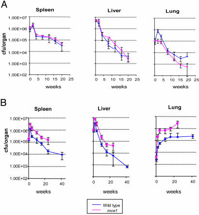
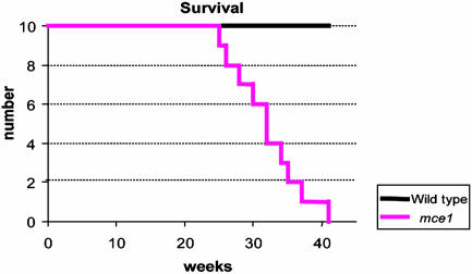
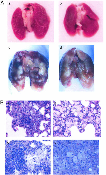
An estimated one-third of the world's population is latently infected with Mycobacterium tuberculosis, the etiologic agent of tuberculosis. Here, we demonstrate that, unlike wild-type M. tuberculosis, a strain of M. tuberculosis disrupted in the mce1 operon was unable to enter a stable persistent state of infection in mouse lungs. Instead, the mutant continued to replicate and killed the mice more rapidly than did the wild-type strain. Histological examination of mouse lungs infected with the mutant strain revealed diffusely organized granulomas with aberrant inflammatory cell migration. Murine macrophages infected ex vivo with the mutant strain were reduced in their ability to produce tumor necrosis factor alpha, IL-6, monocyte chemoattractant protein 1, and nitric oxide (NO), but not IL-4. The mce1 mutant strain complemented with the mce1 genes stimulated tumor necrosis factor alpha and NO production by murine macrophages at levels stimulated by the wild-type strain. These observations indicate that the mce1 operon mutant is unable to stimulate T helper 1-type immunity in mice. The hypervirulence of the mutant strain may have resulted from its inability to stimulate a proinflammatory response that would otherwise induce organized granuloma formation and control the infection without killing the organism. The mce1 operon of M. tuberculosis may be involved in modulating the host inflammatory response in such a way that the bacterium can enter a persistent state without being eliminated or causing disease in the host.


Figures

Fig. 1.
Fig. 1.
(A) mce1 operon based on M. tuberculosis H37Rv genome sequence (8). (B) Western blot analysis of wild-type M. tuberculosis H37Rv (lane 1) (Rv 0169), yrbE1B (Rv0168) H37Rv mutant (lane 2), and mce1A Erdman mutant (lane 3). The immunoblot analysis was performed with a rabbit polyclonal antibody raised against Mce1A, Mce1C, Mce1E, or Mce1F. The antibodies recognize the respective mce1 operon proteins encoded by the wild-type strain only. Disruption of yrbE1B or mce1A by the hygromycin resistance gene cassette led to loss of expression of downstream genes in the operon.
Fig. 2.
Fig. 2.
Time course of infection after tail vein injection of bacillus Calmette-Guérin wild-type and mce1 mutant (A) and M. tuberculosis Erdman wild-type and mce1 mutant (B). Recovery of the bacteria is enumerated by cfu per organ at 0, 2, 4, 10, 16, 25, and 41 wk after injection. The curve for cfu recovered from mice infected with the M. tuberculosis mce1 mutant stops at ≈27 wk, because all of the initially infected mice were dead by this time point.
Fig. 3.
Fig. 3.
Survival kinetics of BALB/c mice infected with M. tuberculosis Erdman. Ten mice each were initially infected via tail vein and followed for the indicated number of weeks. By 41 wk, all mice infected with the mce1 mutant strain were dead.
Fig. 4.
Fig. 4.
(A) Gross examination of lungs of BALB/c mice infected with bacillus Calmette-Guérin or M. tuberculosis. Lungs of mice were examined after 16 wk of infection with the wild-type bacillus Calmette-Guérin (a) or bacillus Calmette-Guérin mce1 mutant (b). Lungs of mice were examined after 32 wk of infection with wild-type M. tuberculosis Erdman (c) or M. tuberculosis Erdman mce1 mutant (d). Lung lesions with discrete well circumscribed borders are evident in wild-type-infected lungs, whereas lungs infected with either of the mce1 mutants show coalescing poorly circumscribed lesions. (B) Histological examination (hematoxylin/eosin stain) of lung sections of BALB/c mice infected with bacillus Calmette-Guérin or M. tuberculosis Erdman. At 12 wk of infection, there is greater and denser lymphocyte infiltration in lungs infected with the wild-type bacillus Calmette-Guérin strain (a), whereas the lungs infected with the mutant bacillus Calmette-Guérin show many foamy macrophages with sparse migration of lymphocytes (b). At 16 wk of infection with M. tuberculosis, lungs infected with the wild-type strain show densely packed lymphocytes surrounded by macrophages (c), whereas lungs infected with the mutant strain show mostly macrophages with diffuse distribution of the lymphocyte population (d). (Magnification in B, ×400.)
Fig. 5.
Fig. 5.
TNFα determinations in BALB/c peritoneal macrophages infected ex vivo with bacillus Calmette-Guérin or M. tuberculosis. TNFα levels were measured in cells infected with live bacillus Calmette-Guérin wild-type or mce1 mutant (A), M. tuberculosis Erdman or mce1 mutant (B), and M. tuberculosis wild-type H37Rv or mce1 mutant (C). Control cells were mock-infected with PBS in each experiment.
Fig. 6.
Fig. 6.
Kinetic analysis of TNFα (A), IL-6 (B), and MCP-1 (C) production by RAW macrophages infected with H37Rv wild-type (green) or mce1 mutant (blue) strains at a moi of 1:1. Controls included uninfected RAW cells (maroon) or lipopolysaccharide-treated cells (red).
Fig. 7.
Fig. 7.
(A) Kinetic analysis of nitrite production by RAW macrophages infected with H37Rv wild-type (green) or mce1 mutant (blue) strains at a moi of 1:1. Controls included uninfected RAWs (solid squares) or lipopolysaccharide-treated cells (red). (B) Nitrite production by untreated (black bar) and IFN-γ-pretreated (maroon bar) RAW macrophages infected with H37Rv wild-type or mce1 mutant strains (moi 1:1) 24 h after infection.
Fig. 8.
Fig. 8.
TNFα (A) and nitrite (B) production by IFN-γ-treated murine bone marrow-derived macrophages infected with H37Rv wild-type, mutant, or mce1 operon-complemented strain of M. tuberculosis. The cells were infected with 1 × 10⁵ bacilli at a moi of 1:10, and the TNFα or nitrite measurements were made at 24 h of infection. Each bar represents a mean value ± SD of infection done in triplicate per strain.

References

    1. Parrish, N. M., Dick, J. D. & Bishai, W. R. (1998) Trends Microbiol. 6, 107-112. - PubMed
    1. Mckinney, J. D., zu Bentrup, D. H., Munoz-Elias, E. J., Miczak, A. Cehn, B., Chan, W., Swenson, D., Sacchettini, J. C., Jacobs, W. R. & Russell, D. G. (2000) Nature 406, 735-738. - PubMed
    1. Glickman, M. S., Cox, J. S. & Jacobs, W. R. (2000) Mol. Cell 5, 717-727. - PubMed
    1. Zahrt, T. C. & Deretic, V. (2001) Proc. Natl. Acad. Sci. USA 98, 12706-12711. - PMC - PubMed
    1. Arruda, S., Bomfim, G., Knights, R., Huima-Byron, T. & Riley, L. W. (1993) Science 261, 1454-1457. - PubMed

Publication types

Substances