. 2003 Dec 23;100(26):15625-30.
doi: 10.1073/pnas.0307226100. Epub 2003 Dec 8.

Marker succession during the development of keratinocytes from cultured human embryonic stem cells

Affiliations

Marker succession during the development of keratinocytes from cultured human embryonic stem cells

Howard Green et al. Proc Natl Acad Sci U S A. .

Abstract

Human embryonic stem cells injected into scid mice produce nodules containing differentiated somatic tissues. From the trypsinized cells of such a nodule, we have recovered keratinocytes that can be grown in cell culture. The method of recovery is sensitive enough to detect small numbers of keratinocytes formed in the nodule, but for purposes of analysis, it is preferable to study the development of the entire keratinocyte lineage in culture. The principle of our analysis is the successive appearance of markers, including transcription factors with considerable specificity for the keratinocyte (p63 and basonuclin) and differentiation markers characteristic of its final state (keratin 14 and involucrin). We have determined the order of marker succession during the time- and migration-dependent development of keratinocytes from single embryoid bodies in cell culture. Of the markers we have examined, p63 was the earliest to appear in the keratinocyte lineage. The successive accumulation of later markers provides increasing certainty of emergence of the definitive keratinocyte.


Figures

Fig. 1.
Fig. 1.
Part of a keratinocyte colony formed with 3T3 support from cells of an ES cell-produced nodule in a scid mouse. A large nodule resulting from ES cells injected into the leg muscle of a scid mouse was excised, minced, and trypsin-disaggregated. Cells from the second trypsinization were plated on 3T3 feeders and fed with FAD medium. Eleven days later, a colony with morphology typical of keratinocytes was seen under phase microscopy (A). The colony was fixed and stained for p63 (red), basonuclin (blue), and K14 (green), and the resulting stains were merged (B). Nuclear p63, basonuclin, and cytoplasmic K14 are evident. In separate images for each staining, the two transcription factors are detectable in almost all cells, although not with identical intensity. K14 is present everywhere but is particularly marked at the expanding perimeter of the colony.
Fig. 2.
Fig. 2.
Presence of involucrin in a keratinocyte colony derived as in Fig. 1. A primary colony formed with 3T3 support was fixed and stained on day 20. Each cell containing p63 (red) also contains K14 (green) (A). Most regions of the colony contain involucrin (blue) (B). In squamous epithelium, K14 synthesized in the basal layer persists in the suprabasal layers (60) but not in cornified cells. In comparing A and B it is clear that cells brightly stained for K14 do not contain appreciable involucrin (arrowheads), whereas cells containing involucrin are faintly stained for K14 (arrows). Presumably, complete destruction of K14 has not yet taken place.
Fig. 3.
Fig. 3.
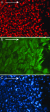
Disappearance of Oct4 from cells migrating out of an embryoid body. A single embryoid body was deposited on a tissue culture dish. Five days later, the culture was fixed and stained for Oct4. Arrows in all photographs indicate direction of migration. The cells of the embryoid body (left) stain brightly for nuclear Oct4 (red). A few cells located in the migration zone close to the embryoid body have retained detectable Oct4, but, beyond this, very few of the 4′,6-diamidino-2-phenylindole-stained nuclei (blue) contain even a trace of Oct4.
Fig. 4.
Fig. 4.
Appearance of p63 and K14 in cells migrating from an attached embryoid body inoculated 15 days previously. The embryoid body is located to the left outside the photograph. Of 517 p63-containing cells, only 30 also contained K14 (green). Because K14 is a cytoplasmic protein, it extends beyond the corresponding p63-containing nucleus. No K14-containing cell lacked p63. Cells lacking both markers are revealed by 4′,6-diamidino-2-phenylindole staining for DNA (blue). Because no supporting feeders are present, those cells, although they lack distinctive morphology, must belong to nonkeratinocyte human lineages.
Fig. 5.
Fig. 5.
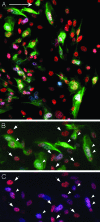
Cells of the keratinocyte lineage close to the migration front. After migration from an embryoid body for 27 days, the migration front (A) shows numerous cells with K14 (green), p63 (red), and basonuclin (blue). At higher power (B and C), the presence of cytoplasmic K14 (B) can be correlated with the presence of purple staining nuclear basonuclin (C, white arrowheads). Other nuclei contain only p63 (white arrows).
Fig. 6.
Fig. 6.
Appearance of involucrin in a stratified keratinocyte colony originating from the migration region of a cultured embryoid body. After 13 days of migration, the cells were trypsinized and inoculated onto 3T3 feeders. Twenty-four days later, a culture containing a colony with the appearance of keratinocytes was fixed and stained. In this merged photograph of the colony, nearly all cells contain p63 (red). Most cells appear to contain K14 (green). Scattered squame-like structures overlying the basal layer contain involucrin (blue).
Fig. 7.
Fig. 7.
Concentration of the keratinocyte lineage at the migration front after the addition of 3T3 cells. After allowing migration from an attached embryoid body for 8 days, 2.6 × 10⁴ irradiated 3T3 cells per cm2 were added to the culture, and incubation was continued for 19 days. The zone close to the migration front is now nearly completely composed of cells containing p63 (red), K14 (green), and basonuclin (blue).
Fig. 8.
Fig. 8.
Marker succession in the keratinocyte lineage.

References

    1. Evans, M. J. & Kaufman, M. H. (1981) Nature 292, 154–156. - PubMed
    1. Martin, G. R. (1981) Proc. Natl. Acad. Sci. USA 78, 7634–7638. - PMC - PubMed
    1. Smith, A. G. (2001) Annu. Rev. Cell Dev. Biol. 17, 435–462. - PubMed
    1. Bagutti, C., Wobus, A. M., Fassler, R. & Watt, F. M. (1996) Dev. Biol. 179, 184–196. - PubMed
    1. Bagutti, C., Hutter, C., Chiquet-Ehrismann, R., Fassler, R. & Watt, F. M. (2001) Dev. Biol. 231, 321–333. - PubMed

Publication types