Review
. 2003 Dec;67(4):593-656.
doi: 10.1128/MMBR.67.4.593-656.2003.

Molecular basis of bacterial outer membrane permeability revisited

Affiliations
Review

Molecular basis of bacterial outer membrane permeability revisited

Hiroshi Nikaido. Microbiol Mol Biol Rev. 2003 Dec.

Abstract

Gram-negative bacteria characteristically are surrounded by an additional membrane layer, the outer membrane. Although outer membrane components often play important roles in the interaction of symbiotic or pathogenic bacteria with their host organisms, the major role of this membrane must usually be to serve as a permeability barrier to prevent the entry of noxious compounds and at the same time to allow the influx of nutrient molecules. This review summarizes the development in the field since our previous review (H. Nikaido and M. Vaara, Microbiol. Rev. 49:1-32, 1985) was published. With the discovery of protein channels, structural knowledge enables us to understand in molecular detail how porins, specific channels, TonB-linked receptors, and other proteins function. We are now beginning to see how the export of large proteins occurs across the outer membrane. With our knowledge of the lipopolysaccharide-phospholipid asymmetric bilayer of the outer membrane, we are finally beginning to understand how this bilayer can retard the entry of lipophilic compounds, owing to our increasing knowledge about the chemistry of lipopolysaccharide from diverse organisms and the way in which lipopolysaccharide structure is modified by environmental conditions.


Figures

FIG. 1.
FIG. 1.
Sequence alignment of porins from α-, β-, and γ-proteobacteria. The classification of the source organisms into γ-, β-, and α-subdivisions is shown at the beginning. Transmembrane β-strands are indicated by highlighting, in red, the presence of hydrophobic amino acid residues at alternate positions. The PEFGGD motif of loop 3 in the Enterobacteriaceae and corresponding sequences are colored in blue. The alignment of E. coli OmpF (ECOOMPF), E. coli OmpC (ECOOMPC), E. coli PhoE (ECOPHOE), Haemophilus influenzae Rd P2 porin (HINP2), Neisseria meningitidis PorB (NMEPORB), Bordetella pertussis porin (BPEPOR), Comamonas acidovorans Omp32 (CACPOR), Rhodopseudomonas blastica porin (RBLPOR), and Rhodobacter capsulatus porin (RCAPOR) is basically that of Jeanteur et al. (303, 304), with minor adjustments. The rest of the sequences (Acidithiobacillus ferrooxidans porin [AFEPOR], Pasteurella multocida OmpH [PMUOMPH], Serratia marcescens OmpF [SMAOMPF], Vibrio cholerae OmpU [VCHOMPU], Photobacterium profundus OmpL [PPROMPL], V. cholerae OmpT [VCHOMPT], and Brucella abortus porin [BABPOR]) were aligned by me. The alignment relied mostly on the plot of (average hydrophobicity + average hydrophobic moment) as specified by Jeanteur et al. (303, 304) and took into account the prediction of turns (485). The Gibbs motif sampling program (442) was also utilized (http://bayesweb.wadsworth.org/gibbs/gibbs.html), although this program predicted only the β-strands facing the lipid bilayer. When multiple sequences were available, deletions and insertions were assumed to have occurred in loops (199); this approach was useful in the analysis of P. multocida OmpH (129, 390) and B. abortus porin (424, 482). The alignment of V. cholerae OmpU, V. cholerae OmpT, and P. profundum OmpL was difficult but was helped by the comparison among these three, as well as with Vibrio fischeri OmpU and VCH1008 from the V. cholerae genome-sequencing project (both sequences retrieved from GenBank). No attempt was made to align the variable-loop sequences carefully.
FIG. 1.
FIG. 1.
Sequence alignment of porins from α-, β-, and γ-proteobacteria. The classification of the source organisms into γ-, β-, and α-subdivisions is shown at the beginning. Transmembrane β-strands are indicated by highlighting, in red, the presence of hydrophobic amino acid residues at alternate positions. The PEFGGD motif of loop 3 in the Enterobacteriaceae and corresponding sequences are colored in blue. The alignment of E. coli OmpF (ECOOMPF), E. coli OmpC (ECOOMPC), E. coli PhoE (ECOPHOE), Haemophilus influenzae Rd P2 porin (HINP2), Neisseria meningitidis PorB (NMEPORB), Bordetella pertussis porin (BPEPOR), Comamonas acidovorans Omp32 (CACPOR), Rhodopseudomonas blastica porin (RBLPOR), and Rhodobacter capsulatus porin (RCAPOR) is basically that of Jeanteur et al. (303, 304), with minor adjustments. The rest of the sequences (Acidithiobacillus ferrooxidans porin [AFEPOR], Pasteurella multocida OmpH [PMUOMPH], Serratia marcescens OmpF [SMAOMPF], Vibrio cholerae OmpU [VCHOMPU], Photobacterium profundus OmpL [PPROMPL], V. cholerae OmpT [VCHOMPT], and Brucella abortus porin [BABPOR]) were aligned by me. The alignment relied mostly on the plot of (average hydrophobicity + average hydrophobic moment) as specified by Jeanteur et al. (303, 304) and took into account the prediction of turns (485). The Gibbs motif sampling program (442) was also utilized (http://bayesweb.wadsworth.org/gibbs/gibbs.html), although this program predicted only the β-strands facing the lipid bilayer. When multiple sequences were available, deletions and insertions were assumed to have occurred in loops (199); this approach was useful in the analysis of P. multocida OmpH (129, 390) and B. abortus porin (424, 482). The alignment of V. cholerae OmpU, V. cholerae OmpT, and P. profundum OmpL was difficult but was helped by the comparison among these three, as well as with Vibrio fischeri OmpU and VCH1008 from the V. cholerae genome-sequencing project (both sequences retrieved from GenBank). No attempt was made to align the variable-loop sequences carefully.
FIG. 2.
FIG. 2.
Structure of the OmpF porin of E. coli. (A) View of the trimer from the top, that is, in a direction perpendicular to the plane of the membrane. Loop 2, colored blue, plays a role in interaction of the monomer with its neighboring unit. Loop 3, colored orange, narrows the channel. (B) View of the monomeric unit from the side, roughly in the direction of the arrow in panel A. Loops 2 and 3 are colored as in panel A. (C) View of the monomeric unit from the top, showing the “eyelet” or the constricted region of the channel. The eyelet is formed by Glu117 and Asp113 from the L3 loop, as well as four basic residues from the opposing barrel wall, Lys16, Arg42, Arg82, and Arg132, all shown as spheres. The diagrams are based on PDB file 2OMF. This figure and Fig. 4 and 6 were drawn by using the program PyMol (Warren L. DeLano, DeLano Scientific LLC, San Carlos, Calif. [http://www.pymol.org]).
FIG. 3.
FIG. 3.
Folding model of OmpA-OprF family slow porins. The major fraction of the population folds as a two-domain protein (left) and is important in binding the OM to the underlying peptidoglycan, since the C-terminal globular domain contains a peptidoglycan-binding motif (165, 342). A minor fraction of the population, however, folds differently to produce an open β-barrel (right). In E. coli, which produces trimeric, high-permeability porins, the presence of this fraction has no functional consequence. However, in fluorescent pseudomonads, which lack the high-permeability porin, this fraction functions as the major nonspecific porin. This fraction also tends to form a loosely associated oligomeric structure, as shown. The oligomer is shown as a trimer only for illustrative purposes. Modified from reference with permission of the publisher.
FIG. 4.
FIG. 4.
X-ray crystallographic structure of LamB. (A) Side view of the monomeric unit. The β-barrel contains 18 strands in this protein, in contrast to the 16 strands seen in the trimeric porins. In addition to loop 3 (orange), loop 1 (red) folds deeply into the channel. Loop 2 (blue) folds outward and interacts with the neighboring subunit in the trimer, as in OmpF (Fig. 2A). Other loops also are often large and tend to cover the entrance of the channel from the outside. (B) View of the monomeric unit from the top. The greasy slides (Tyr41, Tyr6, Trp420, Trp358, and Phe227) are shown as blue stick diagrams, and Tyr118, which constricts the diffusion channel from the opposite side, is shown as a yellow stick diagram. (C) View of the greasy slide and its interaction with maltotriose. This is a side view with the front of the β-barrel cut out for a better view of the slide. The aromatic residues that comprise the greasy slide and Tyr118 are shown as stick diagrams colored as in panel B. The maltotriose molecule (Triose) is shown as a stick diagram colored orange. The coloring of the loops is the same as in panel A. The diagrams are based on PDB coordinate files 1MAL and 1MPN.
FIG. 5.
FIG. 5.
Hypothetical models of the ExbBD-TonB (left) and TolQR-TolB-PAL (right) systems. As seen, most proteins in these two systems are similar in their membrane topology, except for the periplasmic protein To1B and the OM lipoprotein PAL, which do not have counterparts in the Ton system. TonB is drawn as though it will interact directly with the OM receptors while still associated with the inner membrane (IM). However, this may be an oversimplification, and there are pieces of evidence that favor the shuttling of TonB between the OM and the inner membrane (372).
FIG. 6.
FIG. 6.
X-ray crystallographic structures of the ferric citrate receptor, FecA, of E. coli. (A) Side view of the unliganded FecA. The “plug” domain inside the β-barrel is shown in orange. At its N-terminal end, the short sequence comprising the Ton box is shown in light blue. Loops 7 and 8 are shown in deep blue and mauve, respectively. (B) Liganded FecA. On binding of the ferric citrate (with two large blue balls near the top indicating the two iron atoms, and citrate molecules in stick diagrams), large displacements are seen at the N-terminal end of the plug domain, where residues 80 through 95 (including the Ton box of residues 80 to 84) become disordered and invisible. Loops 7 and 8 also undergo large conformational changes, with the loss of part of the helical structure in loop 7. The diagrams are based on PDB files 1KMO and 1KMP.
FIG. 7.
FIG. 7.
Major export pathways of proteins across the OM. Components of type I, type II, and type III secretion pathways are shown schematically. The type I pathway is composed of an ABC transporter (here HlyB for E. coli hemolysin), a membrane fusion protein (MFP) family protein (HlyD), and an OM channel (TolC). In the type II pathway, the proteins reach the periplasmic space via the Sec pathway and are then secreted across the OM by a machinery with many components, including pilin-like proteins (pseudopilins). The proteins are labeled according to the universal Gsp (general secretory pathway) nomenclature, and the scheme owes most to a recent review by Thanassi (652). The energy is apparently supplied by ATP hydrolysis by the GspE protein. The type III system is involved in the secretion (or perhaps injection) of virulence-related proteins into animal and plant host cells. Many of the components have homologies to the proteins of the flagellar hook and basal plate system. The figure is based mainly on a recent proposal that relies heavily on this similarity (73), as well as experimental studies of the “needle complex” (354, 648). The names of the proteins are those from the type III pathway in S. enterica serovar Typhimurium, with those from that of Y. enterocolitica shown in parentheses. The energy for export is thought to be supplied by the InvC (YscN) ATPase. IM, inner membrane.
FIG. 8.
FIG. 8.
X-ray crystallographic structures of the TolC trimer and the OM-puncturing needle apparatus of phage T4. (A) TolC trimer. Each subunit is shown in a different color. At the top, a 12-strand β-barrel is formed by each subunit contributing four strands each. Below the barrel, there is a long periplasmic tunnel composed of 12 α-helices. Structure Based on PDB coordinate file 1EK9. (B) OM-puncturing needle apparatus of phage T4. This structure, produced by protein gp5, is also trimeric, with each monomer shown in a different color. The extended needle structure, which traverses the OM and the periplasm just like the β-barrel and the α-helical tunnel of TolC, is constructed in a totally different manner, as a trimeric β-helix. The structure rich in α-helix, surrounding the upper middle part of the needle, is the lysozyme domain, which is thought to play a role in puncturing a hole in the peptidoglycan. The domain at the very top interacts with another protein, gp27, which was omitted for clarity. Structure based on PDB coordinate file 1K28. This figure was drawn by using program VMD (282).
FIG. 9.
FIG. 9.
Lipid A (from E. coli K-12) and galactosylceramide from intestinal epithelial cells. Groups that may act as H-bond donors are shown in boldface.
FIG. 10.
FIG. 10.
Structure of the R-core oligosaccharide in E. coli K-12 (A) (735) (the Hep residue indicated by the asterisk is replaced by GlcNAc in S. enterica serovar Typhimurium) and in P. aeruginosa (B) (568). Individual LPS molecules may not necessarily contain all of the residues shown. Furthermore, the core structure in the O-antigen-containing LPS of P. aeruginosa is modified from the structure in the R-mutant LPS shown (569). Abbreviations: Glc, d-glucose; Gal, d-galactose; Hep, l-glycero-d-manno-heptose; Kdo, 3-deoxy-d-manno-oct-2-ulosonic acid; EtN, ethanolamine; Rha, l-rhamnose; GalN, d-galactosamine. The anomeric configurations of Hep and Kdo are always α. In the P. aeruginosa core, there are more formal negative charges (2 from Kdo residues plus about 4.5 from monophosphates, assuming roughly 1.5 negative charges per phosphatomonoester, i.e., a total of 6.5), especially concentrated in the trisubstituted heptose residue (although the negative charge is partially compensated by the presence of one positive charge in N-alanylgalactosamine). The E. coli core carries about 5.5 formal negative charges, but they are present in a more dispersed manner.
FIG. 11.
FIG. 11.
Positions of fatty acyl chains in an energy-minimized model of LPS. The positions of fatty acids linked at the 2, 3, 2′, and 3′ positions, as well as those linked as piggybacked residues on the 2′- and 3′-linked 3-OH-myristoyl residues [these are shown as (2′) and (3′)] of E. coli Re LPS are shown by overlaying the structure of the energy-minimized conformer A described by Obst et al. (469) onto the two-dimensional hexagonal lattice.
FIG. 12.
FIG. 12.
Electrostatic interactions between the E. coli LPS and the FhuA protein. The LPS structure is shown in a ball-and-stick diagram, with the oxygen atoms in the acidic moieties indicated by large red balls. A, B, C, D, E, and F refer to the 1-pyrophosphate of GlcNI (the reducing glucosamine), the 4′-phosphate of GlcNII (the nonreducing glucosamine), KDOI carboxylate, KDOII carboxylate, the HepI 4-pyrophosphate, and the HepII 4-phosphate, respectively. The amino acid residues apparently interacting with these anionic groups are shown as thin green sticks, with the basic nitrogen atoms indicated by blue balls. The amino acid residues making tight interactions are discussed in the text. The numbering of residues here is that of the engineered FhuA, which contains an 11-residue insertion after Pro405. Thus, Lys439 and Lys441 here correspond to Lys428 and Lys430 of the native FhuA, the numbering used in Table 1 of reference . In addition, the following residues make somewhat longer-range interactions: Arg382 with the secondary phosphate of 1-pyrophosphate (4.6 Å); Lys306 with 4′-phosphate (6 to 7 Å); Lys351 and Arg384 with KDOI carboxylate (4.7 and 7 Å); Lys439 with KDOII carboxylate (4.9 Å); and Arg474 and Arg435 with HepI 4-pyrophosphate (4.7 and about 10 Å). The figure was drawn with DS Viewer Pro (Accelrys, San Diego Calif.) using PDB file 1QFG. In addition to these electrostatic interactions, some amino acid residues may be involved in H-bonding interactions, which are not shown.
FIG. 13.
FIG. 13.
Adaptative changes in S. enterica serovar Typhimurium lipid A structure in response to a low-Mg2+ environment. Alterations are highlighted in boldface, and the enzymes involved (LpxO, PagP, and PmrAB-regulated enzymes of 5-aminoarabinose addition) are indicated by large type. LpxO generates the (S)-2-hydroxymyristate moiety, as shown. Stereochemistry was not indicated for most of the other asymmetric centers shown in Fig. 9. An additional modification of lipid A induced by PmrAB is the addition of phosphoethanolamine on 1-phosphate, as discussed in the text.
FIG. 14.
FIG. 14.
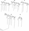
Lipid A with nonclassical structures. (A) E. coli lipid A (as reference). (B) Lipid A from P. aeruginosa (the fatty acid substituents are shorter than in E. coli and are distributed symmetrically over the two glucosamine residues; the “piggyback” fatty acid residues are 2-hydroxylated, so that the lipid A contains more free hydroxyl groups than does the E. coli lipid A; the acyl residue shown at the 3-position may be absent). (C) Lipid A from P. gingivalis (R is H in one strain and an acyl group in another [see the text]). (D) Lipid A from R. leguminosarum (the “reducing” glucosamine residue has been converted into 2-amino-2-deoxygluconic acid; the usual phosphate group at the 4′-position is replaced by a galacturonic acid residue [58], and the characteristic 27-OH-C28:0 acid was found most recently to occur as a piggyback substitutent [40]). (E) Lipid A from Rhodopseudomonas viridis, based on a 2,3-diaminoglucose monomer and totally lacking phosphate substituents (409) (not many details are known about this structure, and the stoichiometry of acyl substituents has not been determined; although the 27-OH-C28:0 acid is known to be present, its position is not known).
FIG. 15.
FIG. 15.
Extractable lipids that replace or supplement LPS in various bacteria. (Left) A sphingolipid, d-glucuronosylceramide, from Sphingomonas yanoikuyae (437). (Center) A sulfonolipid from surface-grown F. johnsoniae (1). (Right) An ornithine lipid from Paracoccus denitrificans (734). In the last lipid, the positions of the double bonds have not been determined and a biosynthetic pathway via cis-vaccenic acid has been assumed.

References

    1. Abbanat, D. R., W. Godchaux III, and E. R. Leadbetter. 1988. Surface-induced synthesis of new sulfonolipids in the gliding bacterium Cytophaga johnsonae. Arch. Microbiol. 149:358-364.
    1. Abbanat, D. R., E. R. Leadbetter, W. Godchaux III, and A. Escher. 1986. Sulfonolipids are molecular determinants of gliding motility. Nature 324:367-369.
    1. Abergel, C., E. Bouveret, J. M. Claverie, K. Brown, A. Rigal, C. Lazdunski, and H. Benedetti. 1999. Structure of the Escherichia coli TolB protein determined by MAD methods at 1.95 Å resolution. Struct. Fold Des. 7:1291-1300. - PubMed
    1. Achouak, W., J. M. Pages, R. De Mot, G. Molle, and T. Heulin. 1998. A major outer membrane protein of Rahnella aquatilis functions as a porin and root adhesion. J. Bacteriol. 180:909-913. - PMC - PubMed
    1. Ackermann, H.-W. 1999. Tailed bacteriophages: The order Caudovirales. Adv. Virus Res. 51:135-201. - PMC - PubMed

Publication types

LinkOut - more resources