Review
. 2003 Dec;67(4):686-723.
doi: 10.1128/MMBR.67.4.686-723.2003.

A continuum of anionic charge: structures and functions of D-alanyl-teichoic acids in gram-positive bacteria

Affiliations
Free PMC article
Review

A continuum of anionic charge: structures and functions of D-alanyl-teichoic acids in gram-positive bacteria

Francis C Neuhaus et al. Microbiol Mol Biol Rev. 2003 Dec.
Free PMC article

Abstract

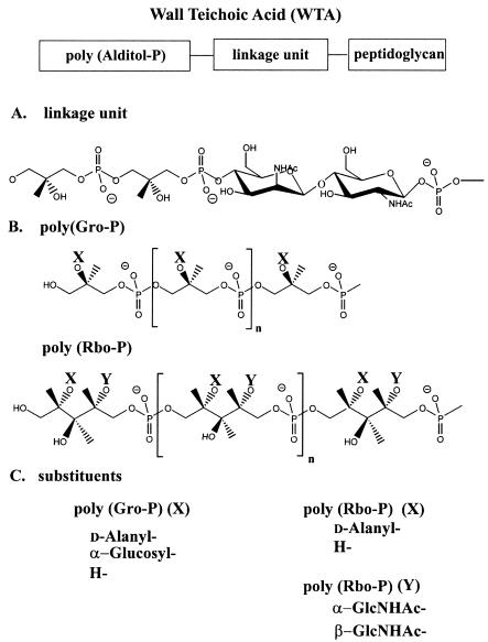
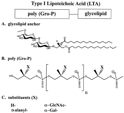
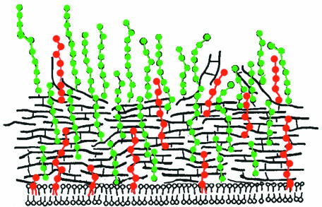
Teichoic acids (TAs) are major wall and membrane components of most gram-positive bacteria. With few exceptions, they are polymers of glycerol-phosphate or ribitol-phosphate to which are attached glycosyl and D-alanyl ester residues. Wall TA is attached to peptidoglycan via a linkage unit, whereas lipoteichoic acid is attached to glycolipid intercalated in the membrane. Together with peptidoglycan, these polymers make up a polyanionic matrix that functions in (i) cation homeostasis; (ii) trafficking of ions, nutrients, proteins, and antibiotics; (iii) regulation of autolysins; and (iv) presentation of envelope proteins. The esterification of TAs with D-alanyl esters provides a means of modulating the net anionic charge, determining the cationic binding capacity, and displaying cations in the wall. This review addresses the structures and functions of D-alanyl-TAs, the D-alanylation system encoded by the dlt operon, and the roles of TAs in cell growth. The importance of dlt in the physiology of many organisms is illustrated by the variety of mutant phenotypes. In addition, advances in our understanding of D-alanyl ester function in virulence and host-mediated responses have been made possible through targeted mutagenesis of dlt. Studies of the mechanism of D-alanylation have identified two potential targets of antibacterial action and provided possible screening reactions for designing novel agents targeted to D-alanyl-TA synthesis.


Figures

FIG. 1.
FIG. 1.
Protonated d-alanyl ester substituent linked to the 2′ hydroxyl of a Gro-P unit (sn-glycerol 1-phosphate). Ion pairing of the phosphodiester with the protonated amino group occurs on rotation of the phosphodiester linkage.
FIG. 2.
FIG. 2.
WTA. (A) Linkage unit. (B) Poly(Gro-P)(sn-glycerol 3-P) moiety from B. subtilis 168 and poly(Rbo-P) from S. aureus H. (C) Substituents on poly(Rbo-P) and poly(Gro-P) characteristic of these bacteria.
FIG. 3.
FIG. 3.
Type I LTA. (A) Glycolipid anchor. (B) Poly(Gro-P) (sn-glycerol 1-P). (C) Substituents (X).
FIG. 4.
FIG. 4.
Assembly of the glucosylated poly(Gro-P) WTA of B. subtilis 168. TagD, TagE, and TagF are enzymes in WTA synthesis, and TagO, TagA, and TagB participate in linkage unit synthesis. Reprinted from reference and amended with permission of the authors and publisher.
FIG. 5.
FIG. 5.
Topography of WTA and LTA in S. aureus. Peptidoglycan (black lines) and WTA (green symbols) adapted with permission of the author and publisher of reference . Topology of LTA (red symbols) and WTA derived from references , , , , and .
FIG. 6.
FIG. 6.
Formation of the acyl phosphodiester intermediate. Stabilization by hydrogen bonding of the C-2 hydroxyl of glycerol increases the electrophilicity of the carbonyl carbon. Protonation of the d-alanyl ester would result in ion pair formation with the phosphodiester and thus would inhibit the formation of the intermediate.
FIG. 7.
FIG. 7.
(A) Enhanced electrophilicity of the carbonyl carbon in d-alanyl-glycerol by hydrogen bonding to the C-1 hydroxyl of glycerol. (B) Migration of the d-alanyl ester via the cyclic ortho ester intermediate. These structures are based on those proposed for acyl mobility and reactivity in aminoacyl-tRNA (75, 192, 504).
FIG. 8.
FIG. 8.
Predicted conformations of the d-alanyl ester on (Gro-P)2Gro. Two conformations are shown: up-chain ion pairing (A) and down-chain ion pairing (B). In panel A, the Nformula image
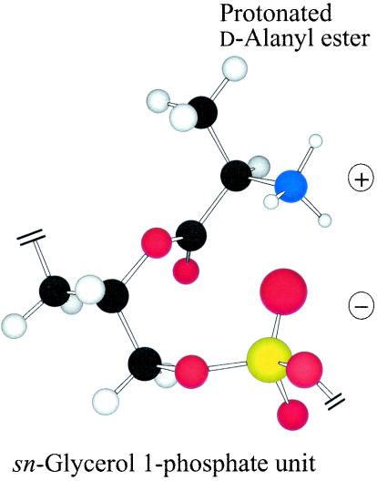
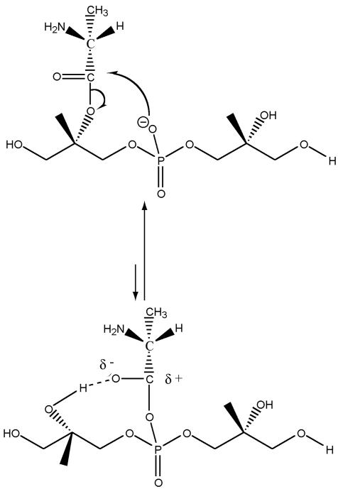
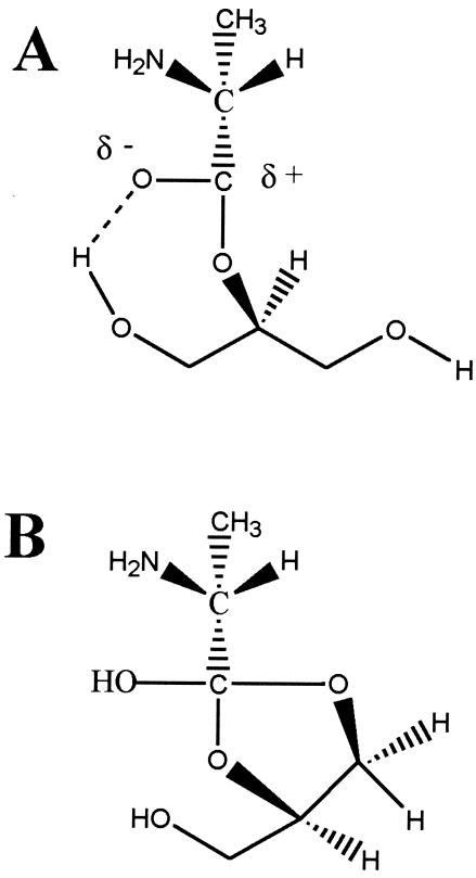
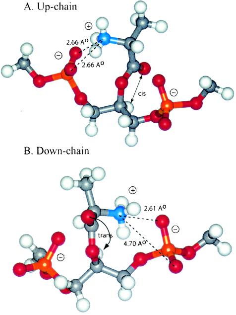
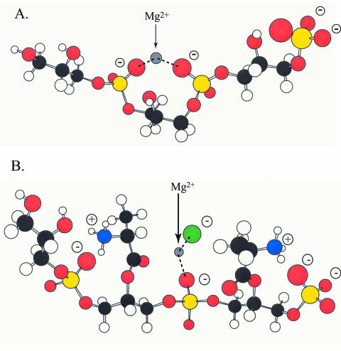
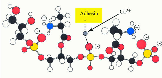
O distances are both 2.66 Å. In panel B, the corresponding distances are 2.61 and 4.70 Å. In panel A, the carbonyl oxygen and the C-2 proton of glycerol are cis, and in panel B they are trans. Resonance stabilization in the ester linkage determines a rotational barrier (495) between the two conformers. For this figure, the flanking glycerol residues are truncated. The conformations were calculated by the semi-empirical molecular orbital method, MNDO-PM3 (, ; Arnold and Neuhaus, unpublished).
FIG. 9.
FIG. 9.
Comparison of the dlt operons from L. rhamnosus, B. subtilis 168, and S. agalactiae. The accession numbers are AF192553 (U43894), X73124, and AJ291784, respectively. In addition, the sequences for dlt from L. monocytogenes (AJ012255), S. mutans (AF051356; AF049357), S. aureus (AF101234; D86240), S. pneumoniae R6 (AE008562), L. lactis (AE006358), S. xylosus (AF032440), S. pyogenes (AE004092), S. gordonii DL1 subsp. Challis (AF059609), and L. plantarum (NC_004567) have been determined. For alignment and comparison of the dlt proteins, the http://genolist.pasteur.fr/SubtiList site is invaluable. Each of the red genes is common to all dlt operons. The green genes in S. agalactiae represent a novel two-component regulatory system (405). The genes in black are not required for d-alanylation. •  is the rho-independent terminator.
FIG. 10.
FIG. 10.
Model for the incorporation of d-alanyl ester residues into membrane-associated LTA. DltD provides binding sites for Dcp and Dcl on the cytoplasmic leaflet. DltB provides a putative channel for the secretion of d-alanyl-Dcp to the periplasm where d-alanylation occurs.
FIG. 11.
FIG. 11.
Ribbon diagram of the minimized average structure of apo-Dcp (PDP entry 1HQB). Residues shown in white bury the Trp67 side chain (purple) in the hydrophobic core. Other key residues include the conserved Glu33 and Asp38(red) and Ser39 (yellow), as well as a cluster of basic residues (blue) proximal to the phosphopantetheine attachment site (Arg64, Lys65, and Lys72). Reprinted from reference with permission of the publisher.
FIG. 12.
FIG. 12.
Putative binding site for the poly(Gro-P) moiety on apo-Dcp. Surface representations, colored according to electrostatic potential, are shown for apo-Dcp (A) and AcpP (PDP entry 1ACP, model 1) (B). Arg64 is conserved in all Dcp proteins, whereas Lys65 in L. rhamnosus Dcp is not conserved. Reprinted from reference with permission of the publisher.
FIG. 13.
FIG. 13.
Proposed mechanism for the formation of membrane-associated d-alanyl-LTA from d-alanyl-Dcp. B·· indicates an unknown proton acceptor for generating nucleophile. The electrostatic interaction between d-alanyl-Dcp and the phosphodiester anion may be due to Arg64. Reprinted from reference with permission.
FIG. 14.
FIG. 14.
Effect of Dcp concentration on the formation of d-Alanyl-Dcp from membrane-associated d-alanyl-LTA. The reaction mixture contained 20 μg of membrane-associated d-[14C]alanyl-LTA and the indicated amounts of Dcp or ACP in 15 μl of reaction mixture. In mixtures containing dltD::cat membranes, dltD was insertionally inactivated (118). The amounts of d-[14C]alanyl-Dcp formed were quantitated by nondenaturing polyacrylamide gel electrophoresis by the method of Heaton and Neuhaus (211). Reproduced from reference with permission.
FIG. 15.
FIG. 15.
Transacylation of d-alanyl ester residues along and among the chains of LTA and WTA. Interchain and intrachain transacylation is illustrated as a mechanism for distributing the esters and for the formation of d-alanyl-WTA from d-alanyl-LTA. Whether this process occurs by the mechanism described in reference or that described in reference has not been determined. A+ represents the d-alanyl ester.
FIG. 16.
FIG. 16.
Bidentate (A) and monodentate (B) binding of the Mg2+ cation by phosphodiester linkages. In panel B, binding to the Cl counterion would occur. For these structures, the ionic radius of nonhydrated Mg2+ is used. Geometry optimization of structures was performed in Chem3D (Molecular Modeling and Analysis), CambridgeSoft.
FIG. 17.
FIG. 17.
Interchain bidentate bridging of TAs by Ca2+. Up-chain and down-chain ion pairings with protonated d-alanyl esters are not illustrated.
FIG. 18.
FIG. 18.
Scheme illustrating the salt-induced transition in the supramolecular organization of the membrane-associated poly(Gro-P) moiety of LTA. At low ionic strength (−NaCl), the poly(Gro-P) chain assumes a stretched conformation, and at high ionic strength (+NaCl), it forms a random coil. Courtesy of T. Gutberlat (presented at the Spring Colloquium on Molecular Modeling, Darmstadt, Germany, 1995) with permission.
FIG. 19.
FIG. 19.
Role of Ca2+ in the presentation of an adhesin. With the exception of adhesin, the structure was energy minimized as described in the legend to Fig. 16B. One or more cations may be involved in the binding of adhesin.
FIG. 20.
FIG. 20.
Continuum of ionic charge. A high-magnification, freeze-substituted image of the septal region of an exponentially growing B. subtilis 168 cell is shown. The tripartite structure of the wall shows the fibrous nature of the outer layer. The electron photomicrograph is reprinted from reference with permission. (A+) represents the d-alanyl esters of TAs, ⊕ represents mobile cations and other fixed cationic functions on peptidoglycan, and ⊖ represents the phosphodiester anionic linkages of TAs and anionic groups of peptidoglycan.

References

    1. Aasjord, P., and A. Grov. 1980. Immunoperoxidase and electron microscopy studies of staphylococcal lipoteichoic acid. Acta Pathol. Microbiol. Scand. Sect. B 88:47-52. - PubMed
    1. Abachin, E., C. Poyart, E. Pellegrini, E. Milohanic, F. Fiedler, P. Berche, and P. Trieu-Cuot. 2002. Formation of d-alanyl-lipoteichoic acid is required for adhesion and virulence of Listeria monocytogenes. Mol. Microbiol. 43:1-14. - PubMed
    1. Alkan, M. L., and E. H. Beachey. 1978. Excretion of lipoteichoic acid by group A streptococci: influence of penicillin on excretion and loss of ability to adhere to human oral epithelial cells. J. Clin. Investig. 61:671-677. - PMC - PubMed
    1. Al-Obeid, S., L. Gutmann, and R. Williamson. 1990. Correlation of penicillin-induced lysis of Enterococcus faecium with saturation of essential penicillin-binding proteins and release of lipoteichoic acid. Antimicrob. Agents Chemother. 34:1901-1907. - PMC - PubMed
    1. Aly, R., and S. Levit. 1987. Adherence of Staphylococcus aureus to squamous epithelium: role of fibronectin and teichoic acid. Rev. Infect. Dis. 9(Suppl. 4):S341-S350. - PubMed

Publication types

MeSH terms

LinkOut - more resources