Review
. 2003;Suppl 37(Suppl ):14-46.
doi: 10.1002/ajpa.10384.

Role of viruses in human evolution

Affiliations
Review

Role of viruses in human evolution

Linda M Van Blerkom. Am J Phys Anthropol. 2003.

Abstract

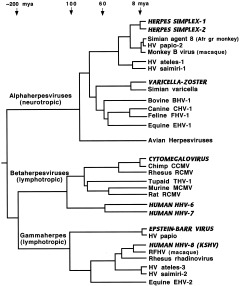
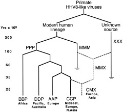
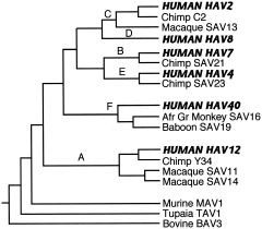
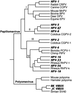
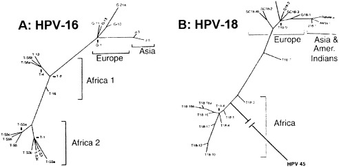
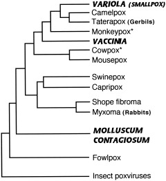
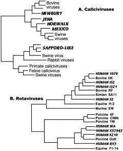
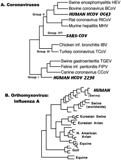
The study of viral molecular genetics has produced a considerable body of research into the sequences and phylogenetic relationships of human and animal viruses. A review of this literature suggests that humans have been afflicted by viruses throughout their evolutionary history, although the number and types have changed. Some viruses show evidence of long-standing intimate relationship and cospeciation with hominids, while others are more recently acquired from other species, including African monkeys and apes while our line was evolving in that continent, and domesticated animals and rodents since the Neolithic. Viral selection for specific resistance polymorphisms is unlikely, but in conjunction with other parasites, viruses have probably contributed to selection pressure maintaining major histocompatibility complex (MHC) diversity and a strong immune response. They may also have played a role in the loss in our lineage of N-glycolylneuraminic acid (Neu5Gc), a cell-surface receptor for many infectious agents. Shared viruses could have affected hominid species diversity both by promoting divergence and by weeding out less resistant host populations, while viruses carried by humans and other animals migrating out of Africa may have contributed to declines in other populations. Endogenous retroviral insertions since the divergence between humans and chimpanzees were capable of directly affecting hominid evolution through changes in gene expression and development.


Figures

Figure 1
Figure 1
Phylogeny of mammalian herpesviruses. Composite tree based on amino‐acid sequences from eight genes. Viruses that regularly infect humans are indicated by bold amd italicized capitals. KSHV, Kaposi's sarcoma herpesvirus; RFHV, retroperitoneal fibromatosis herpesvirus of macaques; Afr gr, African green. Sources: McGeoch and Davison, 1999a; McGeoch et al., 2000; McGeoch, 2001.
Figure 2
Figure 2
Recombination in human herpesvirus‐8. Capital letters (e.g., PPP, BBP) refer to alleles (ORF‐K1, ORF‐75, ORF‐K15) carried by viral subtypes. PPP, original intact form of HHV‐8. MMM, hypothesized older variant carrying M‐associated ORF‐75 allele. XXX, unknown exotic HHV8‐like virus with highly diverged ORF‐K15 allele. AAP, BBP, CCP, and DDP represent variants with different alleles of ORF‐K1. Dashed lines indicate possible recombination events. Reprinted from Hayward (1999), Fig. 5, with permission from Elsevier.
Figure 5
Figure 5
Phylogeny of primate adenoviruses. Composite tree derived from phylogenies based on analysis of VA RNA and DNA polymerase genes (Kidd et al., 1995; Song et al., 1996). A–F are subgenera of primate adenoviruses; branch points between them were derived from comparison of trees from all regions of genome (Bailey and Mautner, 1994). Viruses that regularly infect humans are indicated by bold and italicized capitals. Genetic distances are not to scale.
Figure 3
Figure 3
Phylogeny of mammalian Papovaviridae. Composite tree constructed from phylogenies in Ong et al. (1993), Van Ranst et al. (1995), Chan et al. (1997), and Shadan and Villareal (1993). Viruses that regularly infect humans are indicated by bold amd italicized capitals. Papilloma subtypes I and II infect cutaneous tissue and include viruses that cause warts and other skin conditions; subtype IV infects mucosal tissue and includes malignant genital papillomaviruses HPV‐16 and ‐18; subtype III contains viruses that infect both types of tissue. Polyoma viruses infect kidney cells.
Figure 4
Figure 4
Intratype diversity in papillomaviruses 16 (A) and 18 (B). Phylogenetic trees of HPV16 and HPV18 variants from patients from Africa (Tanzania), Europe (Germany and Scotland), East Asia (Japan), and America (Mundurucu Indian, Brazil), based on analysis of 364‐bp genomic segments of HPV16 and 321‐bp segments of HPV18. HPV18 reference clone was found in both East Asia and South America. HPV45 is included in HPV18 tree to indicate probable African root. Reprinted from Ong et al. (1993), Fig. 3, with permission from American Society for Microbiology and the authors.
Figure 6
Figure 6
Phylogeny of mammalian poxviruses. Approximate relationships as presented in Blasco (1995), Douglas and Dumbell (1996), and Senkevich et al. (1997). Viruses specifically adapted to humans are indicated by bold and italicized capitals. *Maintained in nature in wild rodents.
Figure 7
Figure 7
Common agents of infant diarrhea. A: Caliciviruses. Calicivirus phylogeny derived from sequence analysis of capsid gene of Sapporo‐like viruses (Jiang et al., 1997) and RNA polymerase gene of Norwalk‐like viruses (van der Poel et al., 2000). B: Rotaviruses. Rotavirus phylogeny based on analysis of VP6 capsid protein (reprinted from Tang et al., 1997, Fig. 2, with permission from Elsevier). Human viruses are indicated in bold and italicized capitals.
Figure 8
Figure 8
Respiratory viruses. A: Coronaviruses. Coronavirus phylogenetic relationships derived from analysis of polymerase gene (Stephenson et al., 1999; Marra et al., 2003; Rota et al., 2003). B: Orthomyxovirus: influenza A. Influenza A phylogeny based on nucleotide sequence of nonstructural (NS) protein (Kawaoka et al., 1998). Viruses that infect humans are indicated in bold and italicized capitals.
Figure 9
Figure 9
Paramyxoviruses. Phylogenetic analysis of N open reading frame (Chua et al., 2000). Human viruses are indicated in bold and italicized capitals.
Figure 10
Figure 10
Picornaviruses. Phylogeny of polymerase protein, composite of trees from Rodrigo and Dopazo (1995), Doherty et al. (1999), and Poyry et al. (1999). Human viruses are indicated in bold and italicized capitals. Six most common genera of picornaviruses are indicated next to branches leading to members of each genus. This nomenclature may not reflect genetic relationships, however; note that genus Rhinovirus maps within Enterovirus phylogeny.
Figure 11
Figure 11
Hepatitis viruses. A: Phylogeny of human and primate hepatitis A viruses, based on a 170 base sequence from VP1/P2A junction. Genotypes IA, IB, II, IIIA, IIIB, and VII cause human hepatitis A infection. IA and IB, endemic African strains; II, single isolate from France; IIIA, endemic in many parts of Asia; IIIB, from Japan; VII, single isolate from Sierra Leone. Genotypes IV, V, and VI are strains found in Old World monkeys in Philippines, Kenya, and Indonesia, respectively. For details, see Robertson et al. (1992) and Robertson (2001). B: Phylogenetic analysis of NS5 region of HCV‐like viruses. From Simmonds (2001), Fig. 5, with permission from Society for General Microbiology. Human viruses are indicated in bold italics.
Figure 12
Figure 12
Hepatitis B viruses. Phylogenetic analysis of representative human and nonhuman HBV full genome sequences (Norder et al., 1996; Verschoor et al., 2001). Human viruses are indicated in bold and italicized capitals.
Figure 13
Figure 13
Primate immunodeficiency viruses. Schematic diagram shows phylogenetic relationships and cross‐species transmission of primate lentiviruses, from a number of analyses (Sharp et al., 1999, 2001; Hahn et al., 2000). HIV, human immunodeficiency virus; SIV, simian immunodeficiency virus; SIVsm, sooty mangabey; SIVmac, macaque; SIVsyk, Sykes' monkey; SIVagmVer, vervet; SIVagmGri, grivet; SIVagmTan, tantalus monkey; SIVmnd, mandrill; SIVcpz, chimpanzee; P.t.s., Pan troglodytes schweinfurthii; P.t.t., P. t. troglodytes.
Figure 14
Figure 14
Primate T‐cell lymphotropic viruses (PTLV). Phylogeny of HTLV/STLV types I and II, constructed from data in Slattery et al. (1999), Vandamme et al. (2000), and Van Dooren et al. (2001). STLVs isolated from nonhuman primates are indicated by bold italics; all others including unlabeled lines are HTLV strains isolated from humans. Note interspersal of Central African HTLV‐I subtypes among branches of STLV‐I.
Figure 15
Figure 15
Tissue‐specific gene expression mediated by HERV insertion: salivary amylase. Enhancer sequences in ERVA1 LTR inserted into 5′ flanking region of pancreatic amylase gene (AMY2) activate cryptic promoter in adjoining γ‐actin pseudogene and result in expression of salivary amylase in parotid gland (AMY1). Insertion estimated at 39 mya in an anthropoid ancestor (Samuelson et al., 1990; Ting et al., 1992).

References

    1. Adams NJ, Prescott LE, Jarvis LM, Lewis JCM, McClure MO, Smith DB, Simmonds P. 1998. Detection in chimpanzees of a novel flavivirus related to GB virus‐C/hepatitis G virus. J Gen Virol 79: 1871–1877. - PubMed
    1. Akopov SB, Nikolaev LG, Khil PP, Lebedev YB, Sverdlov ED. 1998. Long terminal repeats of human endogenous retrovirus K family (HERV‐K) specifically bind host cell nuclear proteins. FEBS Lett 421: 229–233. - PubMed
    1. Amengual B, Whitby JE, King A, Cobo JS, Bourhy H. 1997. Evolution of European bat lyssaviruses. J Gen Virol 78: 2319–2328. - PubMed
    1. Armelagos GJ. 1990. Health and disease in prehistoric populations in transition In: Swedlund AC, Armelagos GJ, editors. Disease in populations in transition. New York: Bergin & Garvey; p 127–144.
    1. Armelagos GJ, Dewey JR. 1978. Evolutionary response to human infectious diseases In: Logan MB, Hunt EE, Sr, editors. Health and the human condition: perspectives in medical anthropology. Belmont, CA: Wadsworth Publishing Co. p 101–107.