. 2003 Dec 23;100(26):15895-900.
doi: 10.1073/pnas.2631433100. Epub 2003 Dec 10.

Leukocyte-inspired biodegradable particles that selectively and avidly adhere to inflamed endothelium in vitro and in vivo

Affiliations

Leukocyte-inspired biodegradable particles that selectively and avidly adhere to inflamed endothelium in vitro and in vivo

Harshad S Sakhalkar et al. Proc Natl Acad Sci U S A. .

Abstract

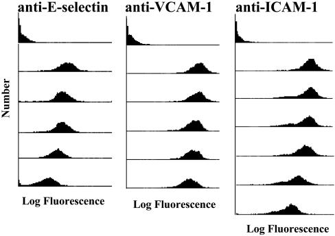
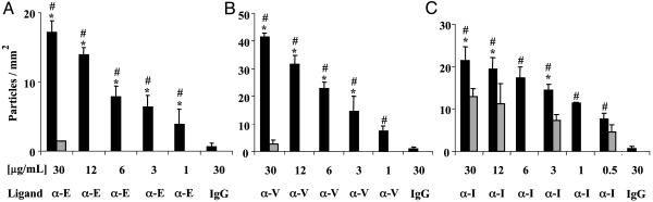
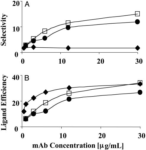
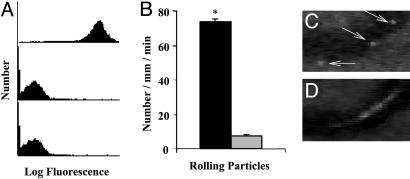
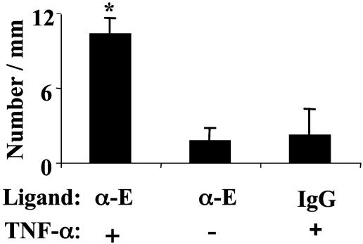
We exploited leukocyte-endothelial cell adhesion chemistry to generate biodegradable particles that exhibit highly selective accumulation on inflamed endothelium in vitro and in vivo. Leukocyte-endothelial cell adhesive particles exhibit up to 15-fold higher adhesion to inflamed endothelium, relative to noninflamed endothelium, under in vitro flow conditions similar to that present in blood vessels, a 6-fold higher adhesion to cytokine inflamed endothelium relative to non-cytokine-treated endothelium in vivo, and a 10-fold enhancement in adhesion to trauma-induced inflamed endothelium in vivo due to the addition of a targeting ligand. The leukocyte-inspired particles have adhesion efficiencies similar to that of leukocytes and were shown to target each of the major inducible endothelial cell adhesion molecules (E-selectin, P-selectin, vascular cell adhesion molecule 1, and intercellular adhesion molecule 1) that are up-regulated at sites of pathological inflammation. The potential for targeted drug delivery to inflamed endothelium has significant implications for the improved treatment of an array of pathologies, including cardiovascular disease, arthritis, inflammatory bowel disease, and cancer.


Figures

Fig. 1.
Fig. 1.
mAbs to E-selectin, VCAM-1, and ICAM-1 can be coupled to PLA–PEG particles. Separate aliquots of PLA–PEG particles, precoupled with neutravidin, were incubated in solutions containing a biotinylated mAb to an ECAM at various concentrations (0, 30, 12, 6, 3, 1, or 0.5 μg/ml mAb from top to bottom, respectively). Subsequently, the particles were treated with a FITC labeled antibody and analyzed with FACS. PLA–PEG particles not treated with the FITC-labeled antibody gave results similar to the top histogram (data not shown). The 0.5 μg/ml concentration was only used for anti-ICAM-1. Results typical of n = 5 experiments. Calibration with Quantum Simply Cellular beads indicated that the 30 μg/ml anti-VCAM-1, anti-ICAM-1, and anti-E-selectin PLA–PEG particles had >200,000 bound ligands.
Fig. 2.
Fig. 2.
LEAPs exhibit selective adhesion to cytokine activated HUVEC. Separate sets of PLA–PEG particles, conjugated with a mAb to an ECAM or mouse IgG (negative control), were perfused over HUVEC in an in vitro flow chamber. The number of LEAPs adherent to the HUVEC was determined after 2.5 min of flow. Black bars indicate adhesion to 4 h cytokine (IL-1β in A and C or TNF-α in B) activated HUVEC. Gray bars indicate adhesion to unactivated HUVEC; μg/ml is the concentration of ligand (either a mAb or mouse IgG) used during the conjugation procedure. Ligand indicates which molecule was coupled to the PLA–PEG particles, a mAb to E-selectin (α-E), VCAM-1 (α-V), ICAM-1 (α-I), or mouse IgG (IgG). Shear stress = 1.5 dynes/cm2; n ≥ 3; ANOVA indicated that the level of adhesion of the LEAPs to cytokine activated HUVEC was a function of the concentration of mAb used in the coupling procedure. *, P < 0.05 compared to LEAPs over unactivated HUVEC (gray bars); #, P < 0.05 compared to 30 μg/ml mouse IgG PLA–PEG particles over 4 h IL-1β or TNF-α-activated HUVEC.
Fig. 3.
Fig. 3.
The selectivity and ligand efficiency of LEAP adhesion to HUVEC. (A) The selectivity, defined as the ratio of the number of LEAPs that adhere to inflamed HUVEC relative to the number of LEAPs that adhere to noninflamed HUVEC, was plotted versus the concentration of mAb used in the conjugation. The selectivity is the highest for targeting VCAM-1 or E-selectin and the lowest for ICAM-1. The selectivity for VCAM-1 and E-selectin was a function of the concentration of anti-VCAM-1 and anti-E-selectin used in the conjugation, whereas the selectivity for ICAM-1 appears to be independent of concentration. (B) The ligand efficiency, defined as the ratio of the number of LEAPs that adhere to inflamed HUVEC relative to the number of IgG PLA–PEG particles that adhere to inflamed HUVEC, was plotted versus the concentration of mAb used in the conjugation. The ligand efficiency was a function of the concentration of mAb and reached a maximum value between 27 and 33. Open square, VCAM-1; filled circle, E-selectin; filled diamond, ICAM-1; shear stress = 1.5 dynes/cm2.
Fig. 4.
Fig. 4.
PSGL-1-conjugated PLA–PEG particles adhere to trauma-activated microvascular endothelium in vivo. (A) PLA–PEG particles, precoupled with either 19.ek.Fc (a recombinant PSGL-1 construct) or ek.Fc (negative control), were treated with a mAb to human PSGL-1 (KPL-1) or human P-selectin (HPDG2/3), washed, treated with an FITC-labeled polyclonal antibody, and analyzed by FACS. (Top) 19.ek.Fc-LEAPs treated with a mAb to PSGL-1 (KPL-1). (Middle) 19.ek.Fc-LEAPs treated with a mAb to P-selectin (isotype-matched control mAb). (Bottom) ek.Fc PLA–PEG particles treated with a mAb to PSGL-1 (KPL-1). Results shown are typical of n = 2 separate experiments. (B) 19.ek.Fc or ek.Fc particles were injected into mice (2 × 10⁷ per mouse), and the number of rolling particles was determined. A significantly greater number of rolling 19.ek.Fc-LEAPs was observed compared to ek.Fc PLA–PEG particles. *, P < 0.05. (C and D) A segment of a postcapillary venule from a typical experiment is shown. (C) Three images were taken 1 s apart and superimposed to generate the composite image. The white sphere (marked by the arrows) is a 19.ek.Fc-LEAP rolling along the wall of the venule. (D) Two images were taken 1/30th of a second apart and superimposed to generate the composite image. The white blur is a PLA–PEG particle(s) not interacting with the vessel wall.
Fig. 5.
Fig. 5.
α-E-LEAPs exhibit selective adhesion to TNF-α inflamed endothelium in vivo. Mice were given an intrascrotal injection of TNF-α or no injection. Approximately 2 h later, α-E-LEAPs or rat IgG PLA–PEG particles (negative control) were injected into the mice (5 × 10⁶ per mouse), and the number of adherent particles was observed in the postcapillary venules of the cremaster muscle. A significantly greater number of α-E-LEAPs were adherent in TNF-α-pretreated mice compared to control mice, and a significantly greater number of α-E-LEAPs were adherent in TNF-α-pretreated mice compared to rat IgG PLA–PEG particles. All adherent particles were firmly adherent (i.e., not rolling). Ligand indicates which molecule was coupled to the PLA–PEG particles, a mAb to murine E-selectin (α-E) or rat IgG (IgG); TNF-α indicates pretreatment of mice with TNF-α 2 h before the experiment (+) or no pretreatment (-); *, P < 0.05 compared to right bars. FACS revealed that the α-E-mAb can be conjugated to PLA–PEG particles (data not shown).

References

    1. Cybulsky, M. I. & Gimbrone, M. A., Jr. (1991) Science 251, 788-791. - PubMed
    1. Soriano, A., Salas, A., Sans, M., Gironella, M., Elena, M., Anderson, D. C., Pique, J. M. & Panes, J. (2000) Lab. Invest. 80, 1541-1551. - PubMed
    1. Jones, S. P., Trocha, S. D., Strange, M. B., Granger, D. N., Kevil, C. G., Bullard, D. C. & Lefer, D. J. (2000) Am. J. Physiol. 279, H2196-H2201. - PubMed
    1. Chapman, P. T., Jamar, F., Keelan, E. T., Peters, A. M. & Haskard, D. O. (1996) Arthritis Rheum. 39, 1371-1375. - PubMed
    1. Schurmann, G. M., Bishop, A. E., Facer, P., Vecchio, M., Lee, J. C., Rampton, D. S. & Polak, J. M. (1995) Gut 36, 411-418. - PMC - PubMed

Publication types

MeSH terms

Substances

LinkOut - more resources