. 2003 Dec 23;100(26):15853-8.
doi: 10.1073/pnas.2136825100. Epub 2003 Dec 10.

Evidence that transgenes encoding components of the Wnt signaling pathway preferentially induce mammary cancers from progenitor cells

Affiliations

Evidence that transgenes encoding components of the Wnt signaling pathway preferentially induce mammary cancers from progenitor cells

Yi Li et al. Proc Natl Acad Sci U S A. .

Abstract

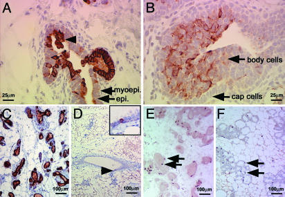
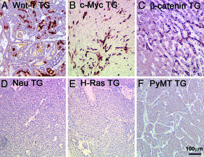
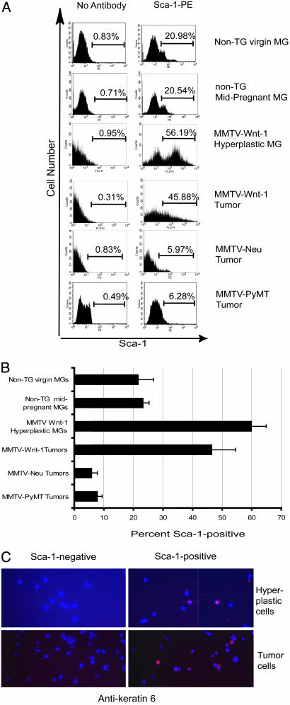
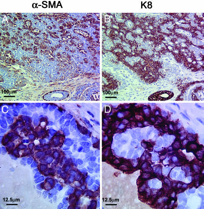
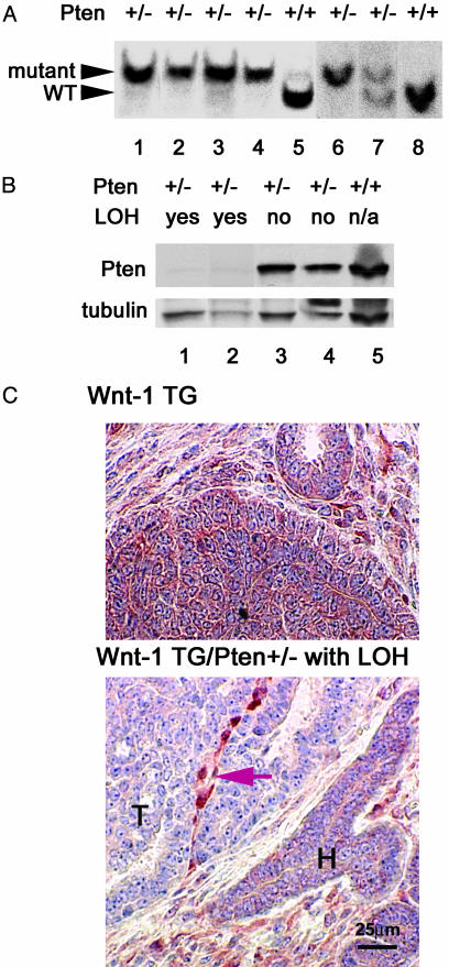
Breast cancer is a genetically and clinically heterogeneous disease, and the contributions of different target cells and different oncogenic mutations to this heterogeneity are not well understood. Here we report that mammary tumors induced by components of the Wnt signaling pathway contain heterogeneous cell types and express early developmental markers, in contrast to tumors induced by other signaling elements. Expression of the Wnt-1 protooncogene in mammary glands of transgenic mice expands a population of epithelial cells expressing progenitor cell markers, keratin 6 and Sca-1; subsequent tumors express these markers and contain luminal epithelial and myoepithelial tumor cells that share a secondary mutation, loss of Pten, implying that they arose from a common progenitor. Mammary tumors arising in transgenic mice expressing beta-catenin and c-Myc, downstream components of the canonical Wnt signaling pathway, also contain a significant proportion of myoepithelial cells and cells expressing keratin 6. Progenitor cell markers and myoepithelial cells, however, are lacking in mammary tumors from transgenic mice expressing Neu, H-Ras, or polyoma middle T antigen. These results suggest that mammary stem cells and/or progenitors to mammary luminal epithelial and myoepithelial cells may be the targets for oncogenesis by Wnt-1 signaling elements. Thus, the developmental heterogeneity of different breast cancers is in part a consequence of differential effects of oncogenes on distinct cell types in the breast.


Figures

Fig. 1.
Fig. 1.
Keratin 6-positive cells are expanded in hyperplastic mammary glands from MMTV-Wnt-1 TG mice. Immunohistochemical staining was used to detect keratin 6 (AD) or BrdUrd (E and F) in mammary gland sections from MMTV-Wnt-1 TG mice that were 3-week-old (A) or 3-month-old (C and E) virgins, from non-TG females that were 3 weeks old (B), 3 months old (D), or in mid-pregnancy (F). The hyperplastic keratin 6-positive cells projecting into the lumen are indicated by an arrowhead (A). The insert in D is a 4-fold higher view of the site denoted with an arrowhead. Staining for BrdUrd, as represented by arrows, demonstrates similar levels of proliferation in mammary glands from MMTV-Wnt-1 TG virgin (E) and wild-type mid-pregnancy (F) mice. Cell types are indicated by arrows in A and B; scale bars are shown in each panel.
Fig. 2.
Fig. 2.
Keratin 6-positive cells in mammary tumors arising in TG mice expressing components of the Wnt signaling pathway. Keratin 6 was detected by immunohistochemical staining of tumors from mice carrying the indicated MMTV TG. The scale is shown in F.
Fig. 3.
Fig. 3.
Increased proportions of Sca-1-positive cells in hyperplastic mammary glands and mammary tumors from MMTV-Wnt-1 TG mice. Representative FACS histograms (A) and a summary bar graph (B) representing data from three to five independent FACS experiments are shown for mammary glands from wild-type virgin and mid-pregnancy mice, hyperplastic mammary glands from MMTV-Wnt-1 TG mice, and mammary tumors from TG mice carrying MMTV-Wnt-1, MMTV-Neu, or MMTV-PyMT transgenes. Hyperplastic cells or tumor cells prepared from MMTV-Wnt-1 TG mice were stained with anti-Sca-1-FITC, sorted by FACS, and stained for keratin 6 (C). Red indicates positive staining for keratin 6. Images were captured by using a ×40 objective.
Fig. 4.
Fig. 4.
Abnormal myoepithelial cells in mammary tumors from MMTV-Wnt-1 TG mice. Consecutive paraffin sections of a mammary tumor from an MMTV-Wnt-1 TG mouse were stained for α-SMA and keratin 8 as indicated. Note the large nuclei and disorganization of the α-SMA-positive cells in the tumor (C). The scales are shown in each panel.
Fig. 5.
Fig. 5.
Loss of Pten heterozygosity in both luminal epithelial and myoepithelial cells of mammary tumors from Pten+/-, MMTV-Wnt-1 TG mice. (A) Southern blotting analysis for LOH at the Pten locus in mammary tumors from MMTV-Wnt-1 TG mice; the Pten genotypes are indicated above the panel. Fragments of the wild-type (WT) and target mutant alleles of Pten are indicated by arrowheads. The image is a composite of two independent blots (lanes 1–5 and lanes 6–8). (B) Western blotting analysis for Pten in MMTV-Wnt-1-derived mammary tumors; the Pten genotype and LOH status are indicated above the panel. The image of the same blot after a subsequent incubation with antibodies against γ-tubulin is also shown to indicate variations in amounts of protein loaded. The faint signal in the samples with evidence of LOH is most likely due to protein from the stroma. (C) Immunohistochemical staining for Pten in mammary tumors; the genotype is indicated above each panel. Areas of tumor (T) and hyperplastic (H) ducts are indicated. The string of positively stained cells (indicated by an arrow) probably represents stroma recruited into the tumor. The scale is shown in Lower.

References

    1. Morrison, B. W. & Leder, P. (1994) Oncogene 9, 3417-3426. - PubMed
    1. Rosner, A., Miyoshi, K., Landesman-Bollag, E., Xu, X., Seldin, D. C., Moser, A. R., MacLeod, C. L., Shyamala, G., Gillgrass, A. E. & Cardiff, R. D. (2002) Am. J. Pathol. 161, 1087-1097. - PMC - PubMed
    1. Cardiff, R. D., Anver, M. R., Gusterson, B. A., Hennighausen, L., Jensen, R. A., Merino, M. J., Rehm, S., Russo, J., Tavassoli, F. A., Wakefield, L. M., et al. (2000) Oncogene 19, 968-988. - PubMed
    1. Cardiff, R. D., Sinn, E., Muller, W. & Leder, P. (1991) Am. J. Pathol. 139, 495-501. - PMC - PubMed
    1. Hedenfalk, I., Duggan, D., Chen, Y., Radmacher, M., Bittner, M., Simon, R., Meltzer, P., Gusterson, B., Esteller, M., Kallioniemi, O. P., et al. (2001) N. Engl. J. Med. 344, 539-548. - PubMed

Publication types