. 2004 Feb;15(2):908-21.
doi: 10.1091/mbc.e03-07-0546. Epub 2003 Dec 10.

A striking quality control subcompartment in Saccharomyces cerevisiae: the endoplasmic reticulum-associated compartment

Affiliations

A striking quality control subcompartment in Saccharomyces cerevisiae: the endoplasmic reticulum-associated compartment

Gregory Huyer et al. Mol Biol Cell. 2004 Feb.

Abstract

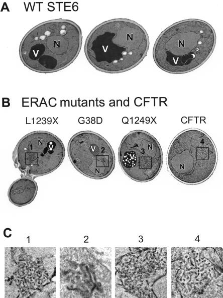
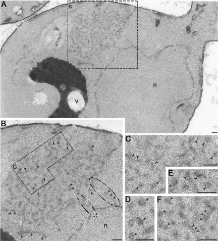
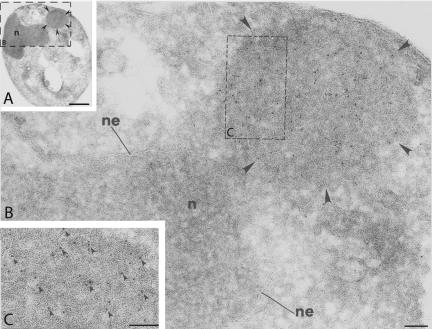
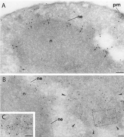
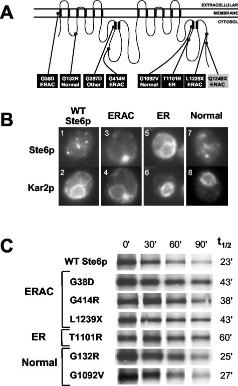
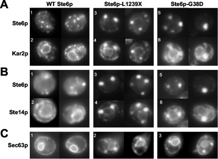
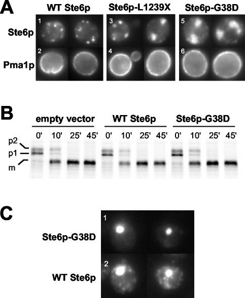
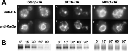
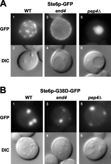
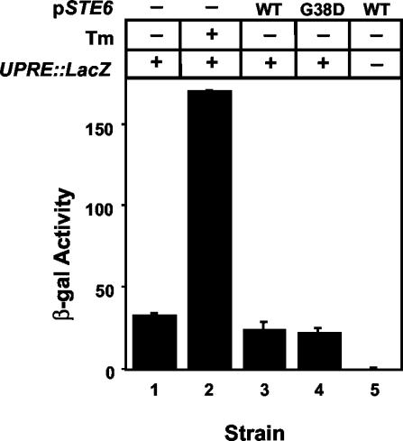
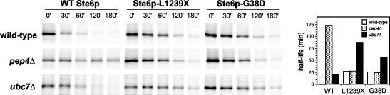
The folding of nascent secretory and membrane proteins is monitored by the endoplasmic reticulum (ER) quality control system. Misfolded proteins are retained in the ER and can be removed by ER-associated degradation. As a model for the ER quality control of multispanning membrane proteins in yeast, we have been studying mutant forms of Ste6p. Here, we identify mislocalized mutant forms of Ste6p that induce the formation of, and localize to, prominent structures that are absent in normal cells. We have named these structures ER-associated compartments (ERACs), based on their juxtaposition to and connection with the ER, as observed by fluorescence and electron microscopy. ERACs comprise a network of tubulo-vesicular structures that seem to represent proliferated ER membranes. Resident ER lumenal and membrane proteins are present in ERACs in addition to their normal ER localization, suggesting there is no barrier for their entry into ERACs. However, the forms of Ste6p in ERACs are excluded from the ER and do not enter the secretory pathway; instead, they are ultimately targeted for ER-associated degradation. The presence of ERACs does not adversely affect secretory protein traffic through the ER and does not lead to induction of the unfolded protein response. We propose that ERACs may be holding sites to which misfolded membrane proteins are specifically diverted so as not to interfere with normal cellular functions. We discuss the likelihood that related ER membrane proliferations that form in response to certain other mutant or unassembled membrane proteins may be substantially similar to ERACs.


Figures

Figure 5.
Figure 5.
ERACs are distinctive membranous structures as visualized by transmission electron microscopy. Yeast strains were examined by transmission EM by using permanganate fixation to enhance visualization of membranes. (A) Micrographs of three cells expressing wild-type Ste6p. (B) Expression of the Ste6p mutants L1239X, G38D, and Q1249X, or CFTR induces formation of tubulo-vesicular structures that are absent in the wild-type strain. (C) Higher magnifications of the tubulo-vesicular areas boxed in B are shown. Cells were prepared for EM as described in MATERIALS AND METHODS. Strains are SM3220 (WT), SM3207 (L1239X), SM3210 (G38D), SM3317 (Q1249X), and SM3245 (CFTR). N, nucleus; V, vacuole.
Figure 6.
Figure 6.
(A and B) ERACs exhibit direct connections to the ER, as detected by OTO fixation. (A) An ERAC is visible adjacent to the perinuclear ER (boxed) by transmission EM in a yeast cell overexpressing Ste6p-G38D. (B) Magnification of area boxed in A, showing direct connections between the ERAC and the ER (circled). (C-F) ERACs are membranous structures. At increased magnification, the membranous structures that comprise ERACs look like railroad tracks (indicated by arrowheads), characteristic of bilayers. C is an enlargement from B, whereas D-F are from other cells overexpressing Ste6p-G38D. Cells (strain SM3210) were prepared as described in MATERIALS AND METHODS. Bar, 0.1 μm.
Figure 7.
Figure 7.
Ste6p-G38D-HA is concentrated in ERACs, as detected by indirect immunogold labeling of ultrathin cryosections. (A) Electron micrograph of a cell expressing Ste6p-G38DHA, with an ERAC marked by arrowheads (n, nucleus). (B) Higher magnification of the area boxed in A. Ste6p-G38D-HA is labeled with 10-nm gold particles and clusters in an ERAC (marked by arrowheads) adjacent to the nuclear envelope (ne)/ER region of the cell. (C) Further magnification of a portion of the ERAC boxed in B, with some of the gold particles labeling Ste6p-G38D-HA indicated by arrowheads. Cells (strain SM3210) were prepared as described in MATERIALS AND METHODS. Ste6p-G38D-HA was detected with a mouse anti-HA antibody followed by a donkey antimouse secondary antibody conjugated to 10-nm gold particles. Bar, 1 μM (A); 0.1 μm (B and C).
Figure 8.
Figure 8.
Ste6p-G38D-HA colocalizes with Kar2p in ERACs, but not in the ER, in double indirect immunogold labeling. (A) Immunogold labeling of Kar2p in a wild-type cell. Kar2p localizes to the nuclear envelope (ne)/perinuclear ER (arrows), and in some places extends out toward the plasma membrane (pm). (B) In a cell expressing Ste6p-G38D-HA, Kar2p (large gold) maintains its normal ER localization, but it is also found to cluster in an ERAC (marked by arrowheads) with Ste6p-G38D-HA (small gold), whereas Ste6p-G38D-HA is only observed in the ERAC. (C) A higher magnification of the colocalization is shown. Kar2p was labeled with 10-nm (large) gold particles, and Ste6p-G38D-HA with 5-nm (small) gold particles. Arrowheads indicate the small gold (i.e., Ste6p-G38D-HA) in proximity of the large gold (i.e., Kar2p). Cells (strain SM3210) were prepared as in Figure 7. Bar, 0.1 μm.
Figure 1.
Figure 1.
(A) Location of mutations in Ste6p. Schematic showing the identity and positions within Ste6p of the six missense and one nonsense mutants identified in this study (black boxes). A previously identified nonsense mutant (ste6-166, Q1249X) also analyzed here is shown (gray box) (Loayza et al., 1998). The localization patterns of the mutant proteins as determined by immunofluorescence are indicated; “normal” refers to a wild-type localization, whereas “other” indicates an indeterminate localization. The coils in the schematic represent membrane spans of Ste6p and the black rectangles indicate the conserved Walker A and B motifs of the nucleotide binding-fold domains (NBDs). (B) ste6 mutants define three groups based on their localization properties. Examples of the coimmunofluorescence pattern of HA-tagged Ste6p (wild-type and mutants) and Kar2p (as a marker for the ER). Cells were costained with an anti-HA mouse antibody (top) and an anti-Kar2p rabbit antibody (bottom). The punctate localization of wild-type Ste6p (1) represents mainly endosomes, and possibly some Golgi (Kelm et al., 2004). The Ste6p mutant proteins G132R and G1092V also show this “normal” wild-type punctate localization pattern (7). The other Ste6p mutant proteins aberrantly localize to ERACs (G38D, G414R, and L1239X; 3) or the ER (T1101R; 5). Only one example from each group is shown: SM3220 (WT); SM3205 (G414R; ERAC); SM3209 (T1101R; ER); and SM3208 (G132R; normal). (C) Metabolic stability of wild-type Ste6p and Ste6p mutant proteins. Cells were pulse-labeled with35S-Met/Cys for 10 min and the label chased for the indicated times. Ste6p levels were analyzed by immunoprecipitation, SDS-PAGE, and PhosphorImager analysis as described in MATERIALS AND METHODS. Strains used are SM3220 (WT), SM3210 (G38D), SM3205 (G414R), SM3207 (L1239X), SM3209 (T1101R), SM3208 (G132R), and SM3206 (G1092V).
Figure 2.
Figure 2.
ER resident proteins localize to the ER and to ERACs, whereas mutant Ste6p is confined to ERACs and excluded from the ER. (A) The localization pattern of wild-type and mutant forms of Ste6p with Kar2p is shown. For HA-tagged wild-type Ste6p and Ste6p-L1239X, cells were costained with an anti-HA mouse antibody to detect Ste6p and an anti-Kar2p rabbit antibody. For GFP-tagged Ste6p-G38D, Ste6p was detected by direct fluorescence, whereas Kar2p was detected by immunofluorescence as described above. Strains used are SM3220 (1 and 2), SM3207 (3 and 4), and SM3898 (5 and 6). (B) The localization pattern of wild-type and mutant forms of Ste6p with the ER membrane protein Ste14p is shown. Cells were prepared as described above, except that an anti-Ste14p rabbit antibody was used. Strains used are SM3966 (1 and 2), SM3967 (3 and 4), and SM4213 (5 and 6). (C) The fluorescence pattern of Sec63p-GFP is shown in cells expressing wild-type, L1239X, or G38D Ste6p. Strains used are SM4207 (1), SM4208 (2), and SM4209 (3).
Figure 3.
Figure 3.
(A) Localization of the plasma membrane protein Pma1p is not affected in cells expressing ERAC-forming Ste6p mutant proteins. The coimmunofluorescence localization pattern of Pma1p with wild-type and mutant forms of Ste6p is shown. Cells were costained with an anti-HA mouse antibody (top) and an anti-Pma1p rabbit antibody (bottom). Strains used are SM3220 (WT), SM3207 (L1239X), and SM3210 (G38D). (B) CPY trafficking is not perturbed when ERACs are present. The trafficking of CPY was followed by pulse-chase analysis in a strain containing an empty vector and strains expressing either wild-type or G38D Ste6p-GFP under the control of a galactose-inducible promoter. Ste6p expression was induced by overnight incubation with 4% galactose, after which ERACs could be observed in ≥50% of cells in the population by fluorescence microscopy. Cells were pulse labeled with35S-Met/Cys for 10 min and the label chased for the indicated times (minutes). CPY was immunoprecipitated, subjected to SDS-PAGE, and analyzed as described in MATERIALS AND METHODS. Strains used are SM4933 (empty vector), SM4934 (WT Ste6p), and SM4935 (Ste6p-G38D). (C) Wild-type Ste6p localizes to ERACs and to endosomes when coexpressed with Ste6p-G38D. To detect wild-type Ste6p-HA, cells were stained with an anti-HA mouse primary antibody and a Cy3-conjugated anti-mouse secondary antibody. Ste6p-G38D-GFP was visualized by direct fluorescence. There is no bleed-through by the GFP signal from Ste6p-G38D into the Cy3 filter used to detect wild-type Ste6p-HA (our unpublished data). The strain used was SM4256.
Figure 4.
Figure 4.
Human CFTR expressed in yeast induces the formation of and localizes to ERACs, whereas human MDR1 does not. (A) The coimmunofluorescence localization pattern of HA-tagged Ste6p, human CFTR, and human MDR1 with Kar2p is shown. Cells were costained with an anti-HA mouse antibody (top) and an anti-Kar2p rabbit antibody (bottom). (B) The metabolic stability of HA-tagged Ste6p, human CFTR, and human MDR1 was examined by metabolic pulse-chase labeling, immunoprecipitation with anti-HA antibodies, and SDS-PAGE as described in MATERIALS AND METHODS. Strains used are SM3220 (STE6), SM3245 (CFTR), and SM3302 (MDR1).
Figure 9.
Figure 9.
Ste6p-G38D does not exit from ERACs into the secretory pathway. The fluorescence pattern and corresponding Nomarski (differential interference contrast) images are shown for cells expressing Ste6p-GFP or Ste6p-G38D-GFP in wild-type, end4, and pep4Δ strains. (A) Ste6p-GFP localizes in a punctate (endosomal) pattern in a wild-type strain (SM3897, 1 and 2), is trapped at the plasma membrane in an end4 strain (SM3863, 3 and 4), and fills the vacuole in a pep4Δ strain (SM4031, 5 and 6). (B) Ste6p-G38D-GFP localizes exclusively to ERACs in all three strains: wild-type (SM3898, 1 and 2), end4 (SM3954, 3 and 4), and pep4Δ (SM1934/pSM1508, 5 and 6).
Figure 10.
Figure 10.
The UPR is not induced by ERAC-forming Ste6p mutant proteins. Strains transformed with a UPRE-lacZ reporter construct and with either empty PGAL vector (-), PGALSTE6 (WT), or PGALste6-G38D (G38D) were induced overnight in 4% galactose and assayed for β-Gal activity. As a control, cells transformed with the empty PGAL vector were treated with the UPR inducer tunicamycin (Tm) for 6 h. Strains used were SM4933 (1 and 2), SM4934 (3), SM4935 (4), and SM4255/pSM1503 (5).
Figure 11.
Figure 11.
Ste6p-G38D and Ste6p-L1239X undergo ubiquitin-mediated ERAD and not Pep4p-dependent vacuolar degradation. Isogenic wild-type, ubc7Δ, and pep4Δ strains overexpressing HA-tagged wild-type, L1239X, or G38D Ste6p were metabolically labeled and analyzed for Ste6p levels by immunoprecipitation and SDS-PAGE as described in MATERIALS AND METHODS. The bar graph shows the half-lives of Ste6p in the indicated strain backgrounds, calculated as described in MATERIALS AND METHODS. Strains used are wild-type: SM4922, SM4923, SM4924; pep4Δ: SM4925, SM4926, SM4927; and ubc7Δ: SM4928, SM4929, and SM4930.

References

    1. Ambudkar, S.V., Dey, S., Hrycyna, C.A., Ramachandra, M., Pastan, I., and Gottesman, M.M. (1999). Biochemical, cellular, and pharmacological aspects of the multidrug transporter. Annu. Rev. Pharmacol. Toxicol. 39, 361-398. - PubMed
    1. Barlowe, C. (2003). Signals for COPII-dependent export from the ER: what's the ticket out? Trends Cell Biol. 13, 295-300. - PubMed
    1. Becker, F., Block-Alper, L., Nakamura, G., Harada, J., Wittrup, K.D., and Meyer, D.I. (1999). Expression of the 180-kD ribosome receptor induces membrane proliferation and increased secretory activity in yeast. J. Cell Biol. 146, 273-284. - PMC - PubMed
    1. Berkower, C., and Michaelis, S. (1991). Mutational analysis of the yeast a-factor transporter STE6, a member of the ATP binding cassette (ABC) protein superfamily. EMBO J. 10, 3777-3785. - PMC - PubMed
    1. Berkower, C., Taglicht, D., and Michaelis, S. (1996). Functional and physical interactions between partial molecules of STE6, a yeast ATP-binding cassette protein. J. Biol. Chem. 271, 22983-22989. - PubMed

Publication types

MeSH terms

LinkOut - more resources