. 2004 Feb;15(2):815-26.
doi: 10.1091/mbc.e03-06-0413. Epub 2003 Dec 10.

A unique region of RILP distinguishes it from its related proteins in its regulation of lysosomal morphology and interaction with Rab7 and Rab34

Affiliations

A unique region of RILP distinguishes it from its related proteins in its regulation of lysosomal morphology and interaction with Rab7 and Rab34

Tuanlao Wang et al. Mol Biol Cell. 2004 Feb.

Abstract

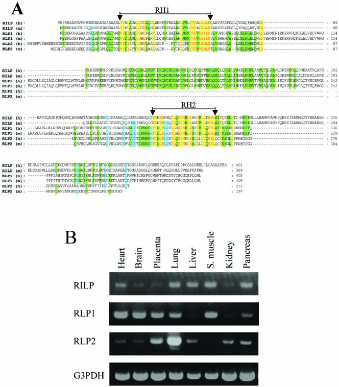
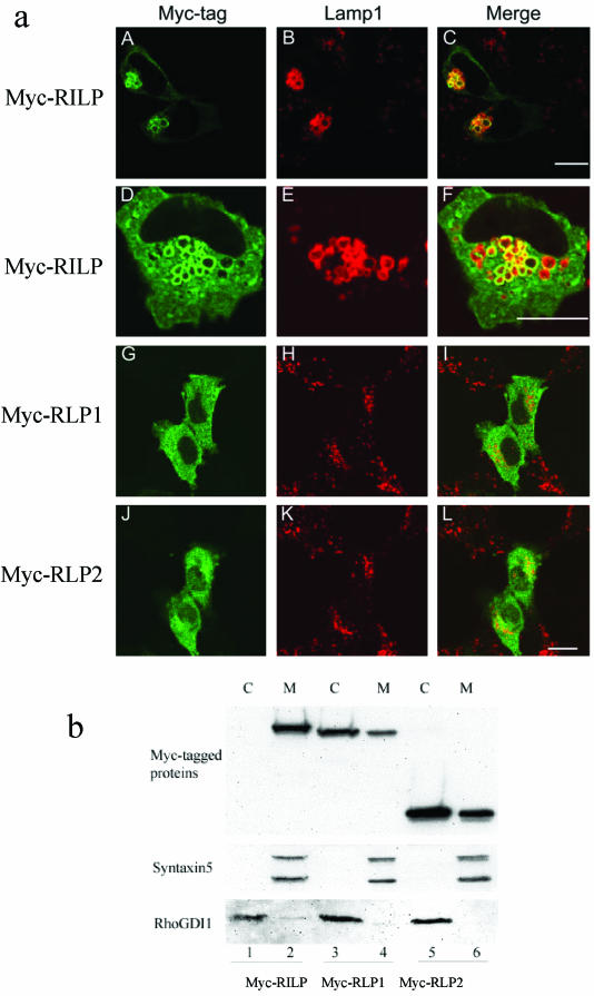
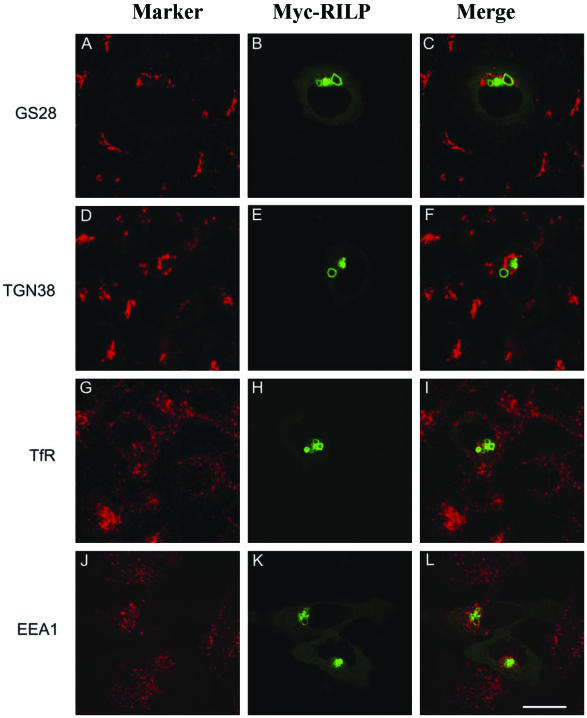
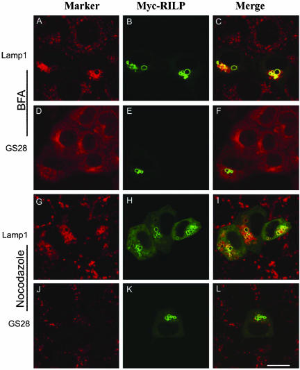
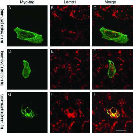
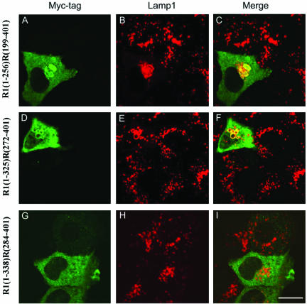
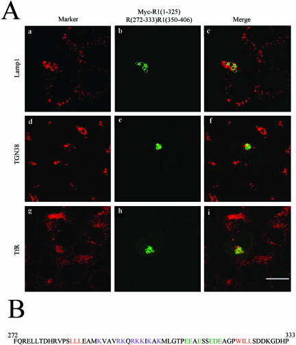
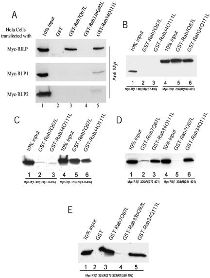
Rab7 and Rab34 are implicated in regulation of lysosomal morphology and they share a common effector referred to as the RILP (Rab-interacting lysosomal protein). Two novel proteins related to RILP were identified and are tentatively referred to as RLP1 and RLP2 (for RILP-like protein 1 and 2, respectively). Overexpression of RILP caused enlarged lysosomes that are positioned more centrally in the cell. However, the morphology and distribution of lysosomes were not affected by overexpression of either RLP1 or RLP2. The molecular basis for the effect of RILP on lysosomes was investigated, leading to the demonstration that a 62-residue region (amino acids 272-333) of RILP is necessary for RILP's role in regulating lysosomal morphology. Remarkably, transferring this 62-residue region unique to RILP into corresponding sites in RLP1 rendered the chimeric protein capable of regulating lysosome morphology. A correlation between the interaction with GTP-bound form of both Rab proteins and the capability of regulating lysosomes was established. These results define a unique region in RILP responsible for its specific role in regulating lysosomal morphology as well as in its interaction with Rab7 and Rab34.


Figures

Figure 1.
Figure 1.
(A) Identification of two novel proteins (RLP1 and RLP2) related to RILP. The amino acid sequences of human (h) and mouse (m) RILP, RLP1, and RLP2 were aligned. The two regions with highest homology were indicated as RH1 (RILP Homology 1) and RH2, respectively. The residues identical or conserved in all proteins were shown in red with a yellow background. Residues identical or conserved in five proteins were shown in blue with a light blue background, while residues identical or conserved in four proteins were shown in black with a green background. (B) RILP, RLP1, and RLP2 are expressed in diverse tissues. The tissue distributions of RILPs were examined by PCR approach using CLONTECH multiple tissue cDNA panels (the PCR product of G3PDH was used for normalization).
Figure 2
Figure 2
(a) RILP but not RLP1 or RLP2 causes enlarged lysosomes positioned centrally in the cell. NRK cells were transfected with constructs for the expression of myc-RILP (A-F), myc-RLP1 (G-I) and myc-RLP2 (J-L). The transfected cells were fixed and double-labeled with polyclonal antibody against the myc tag to reveal the expressed RILP and RLPs (A, D, G, and J) and mAb against rat Lamp1 to reveal lysosomes (B, E, H, and K). RILP colocalizes with lysosomal Lamp1 and causes enlarged lysosomes (A-F), but RLP1 and RLP2 are cytosolic and have no effects on lysosomal morphology (G-L). Bar, 10 μm. (b) RILP is mainly confined to the membrane, while RLP1 and RLP2 are distributed predominantly in the cytosol. Cells expressing Myc-RILP (lanes 1 and 2), Myc-RLP1 (lanes 3 and 4), Myc-RLP2 (lane 5-6) were fractionated into cytosolic (C; lanes 1, 3, and 5) and membrane (M; lanes 2, 4, and 6) fractions, respectively. The fractions were resolved by SDS-PAGE and analyzed by immunoblot to detected the Myc-tagged proteins (top panel) as well as syntaxin5 (serving as membrane protein control) (middle panel) and RhoGDI1 (serving as cytosolic protein control; bottom panel).
Figure 3.
Figure 3.
RILP regulates specifically lysosomes but not Golgi apparatus, early and recycling endosomes. NRK cells (A-I) or Hela cells (J-L) were transfected with myc-RILP expressing construct. The transfected cells were processed for double labeling using antibodies against myc tag and GS28 (A-C), myc tag and TGN38 (D-F), myc tag and transferrin receptor (G-I), or myc tag and EEA1 (J-L). As shown, RILP has no effects on Golgi apparatus, early and recycling endosomes. Note that enlarged lysosomes induced by RILP were not so obvious in HeLa cells, in which lysosomes are collapsed into compact structures. Bar, 10 μm.
Figure 4.
Figure 4.
The effect of RILP on lysosomes is not affected by treatment with brefeldin A (BFA) or nocodazole. NRK cells transfected with myc-RILP expressing construct were treated with BFA (A-F) or nocodazole (G-L) for 1 h at 37°C (both at 10 μg/ml). The cells were then processed for double-labeling using antibodies against myc tag and Lamp1 (A-C and G-I) or myc tag and GS28 (D-F and J-L). As shown, the Golgi apparatus (marked by GS28) was redistributed into ER-like labeling by BFA (D-F) or fragmented by nocodazole treatment (J-L), but the effect of RILP on lysosomes is not affected by either treatments (A-C and G-I). Bar, 10 μm
Figure 5.
Figure 5.
Defining the C-terminal boundary of the RILP region that is responsible for regulating lysosomes. NRK cells were transfected with constructs for expressing myc tagged chimeric proteins consisting of the N-terminal part of RILP (R) and C-terminal part of RLP1 (R1). The transfected cells were then processed for double labeling using antibodies against myc tag and Lamp1. The chimera consisting of N-terminal half of RILP, R(1-198)R1(257-406), or 300-residue N-terminal part of RILP, R(1-300)R1(350-406), had no effects on lysosomes (A-F), but the chimera composed of 333-residue of N-terminal part of RILP, R(1-333)R1(350-406), does (G and H). This defines residue 333 as the C-terminal boundary. Bar, 10 μm
Figure 6.
Figure 6.
Defining the N-terminal boundary of the RILP region that is responsible for regulating lysosomes. NRK cells were transfected with constructs for expressing myc tagged chimeric proteins consisting of N-terminal part of RLP1 (R1) and C-terminal part of RILP (R). The cells were then processed for double labeling using antibodies against myc tag and Lamp1. Chimeric proteins containing C-terminal half of RILP, R1(1-256)R(199-401), (A-C), and C-terminal residues 272-401 of RILP, R1(1-325)R0(272-401), (D-F), affected lysosomes. The C-terminal part of RILP consisting of residues 284-401 lost the ability to affect lysosomes (G-I). This defines residue 272 as the N-terminal boundary of RILP. Bar, 10 μm
Figure 7.
Figure 7.
(A) A unique 62-residue (amino acids 272-333) region of RILP (R1) is sufficient to render RLP1 (R2) capable of regulating lysosomes. NRK cells were transfected with a construct for expressing myc tagged chimera, R1(1-325)R(272-333)R1(350-406). The cells were then processed for double-labeling using antibodies against myc tag and Lamp1 (a-c), myc tag and GS28 (d--f) or myc tag and transferrin receptor (g-i). As shown, residues 272-333 are sufficient for conferring RILP's property to RLP1 in regulating lysosomes. (B) The amino acid sequence of residues 272-333 of RILP. The positive and negative charged residues in the central part were shown in dark pink and dark green, respectively, with the embracing hydrophobic residues marked in red.
Figure 8.
Figure 8.
Correlation between the ability to interact with Rab proteins and regulation of lysosomal morphology. Hela cells were transfected with myc tagged constructs as indicated. The cell lysates were incubated with GST, GST-Rab7Q67L, GST-Rab33bQ92L or GST-Rab34Q111L coupled to GST-Sepharose 4B resin in the presence of 100 μM GTP-γ-S. After extensive washing, the bound proteins were resolved by SDS-PAGE and detected by immunoblotting using antibody against Myc tag. (A) RILP interacts with both Rab7 and Rab34, whereas RLP1 interacts only with Rab34 weakly. The top panel shows that RILP can specifically bind to GST-Rab7Q67L or GST-Rab34Q111L but not GST or GST-Rab33bQ92L. The middle panel indicates RLP1 interacts with Rab34Q111L weakly but not with GST, Rab7Q67L, or Rab33bQ92L. RLP2 did not interact with any Rab protein (bottom panel). (B) C-terminal half of RILP(R1) is responsible for the interaction with Rab7 and Rab34. GST-Rab7Q67L and Rab34Q111L can pull down the chimeric protein R1(1-256)R(199-401) (lanes 4-6) but cannot bind to R(1-198)R1(257-406) (lanes 1-3). (C) Defining C-terminal boundary of the region of RILP (R) responsible for interaction with Rab7 and Rab34. GST-Rab7Q67L and Rab34Q111L can pull down the chimeric protein R(1-333)R1(350-406) (lanes 4-6) but cannot bind to R(1-300)R1(350-406) (lanes 1-3). (D) Defining the N-terminal boundary of the region of RILP (R) responsible for interaction with Rab7 and Rab34. GST-Rab7Q67L and Rab34Q111L can pull down the chimeric protein R1(1-325)R(272-401) (lanes 1-3), whereas R1(1-338)R(284-401) can be pulled down by Rab34Q111L but not Rab7Q67L (lanes 4-6). (E) The 62-residue (amino acids 272-333) region of RILP (R) is sufficient to confer the ability to RLP1(R1) for interaction with Rab7 and Rab34. As shown, efficient retention of Myc-R1(1-325)R(272-333)R1(350-406) by immobilized GST-Rab7Q67L (lane 3) and GST-Rab34Q111L (lane 5) but not GST (lane 2) or GST-Rab33b(Q92L) (lane 4) was observed.

Similar articles

Cited by

References

    1. Barbosa, M.D. et al. (1996). Identification of the homologous beige and Chediak-Higashi syndrome genes. Nature 382, 262-265. - PMC - PubMed
    1. Bucci, C., P. Thomsen, P. Nicoziani, J. McCarthy, and van Deurs, B. (2000). Rab 7, a key to lysosome biogenesis. Mol. Biol. Cell 11, 467-480. - PMC - PubMed
    1. Cantalupo, G., Alifano, P., Roberti, V., Bruni, C.B., and Bucci, C. (2001). Rab-interacting lysosomal protein (RILP): the Rab7 effector required for transport to lysosomes. EMBO J. 20, 683-693. - PMC - PubMed
    1. Caplan, S., Hartnell, L.M., Aguilar, R.C., Naslavsky, N., and Bonifacino, J.S. (2001). Human Vam6p promotes lysosome clustering and fusion in vivo. J. Cell Biol. 154, 109-122. - PMC - PubMed
    1. Chavrier, P., and Goud, B. (1999). The role of ARF and Rab GTPases in membrane transport. Curr. Opin. Cell Biol. 11, 466-475. - PubMed

Publication types

MeSH terms