Review
. 2003 Nov;2(11):891-902.
doi: 10.1038/nrd1227.

Ace revisited: a new target for structure-based drug design

Affiliations
Review

Ace revisited: a new target for structure-based drug design

K Ravi Acharya et al. Nat Rev Drug Discov. 2003 Nov.

Abstract

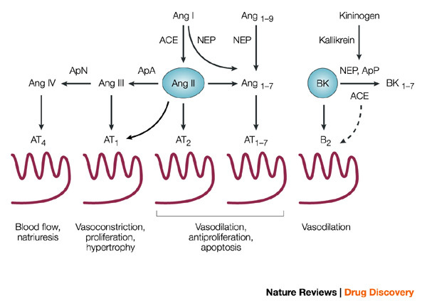
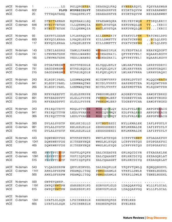
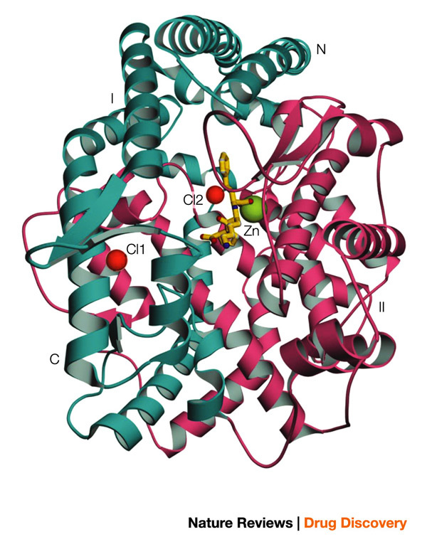
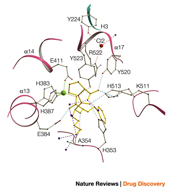
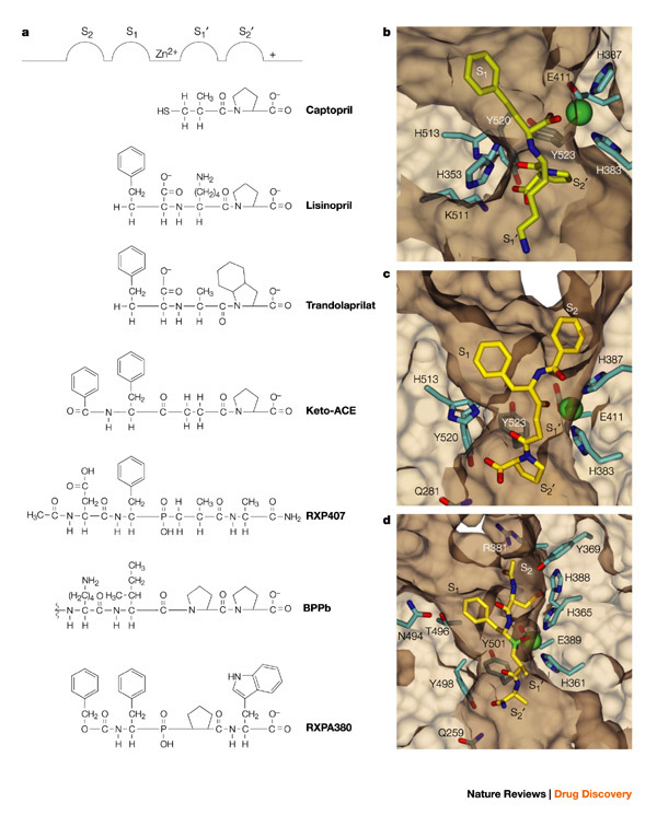
Current-generation angiotensin-converting enzyme (ACE) inhibitors are widely used for cardiovascular diseases, including high blood pressure, heart failure, heart attack and kidney failure, and have combined annual sales in excess of US $6 billion. However, the use of these ACE inhibitors, which were developed in the late 1970s and early 1980s, is hampered by common side effects. Moreover, we now know that ACE actually consists of two parts (called the N- and C-domains) that have different functions. Therefore, the design of specific domain-selective ACE inhibitors is expected to produce next-generation drugs that might be safer and more effective. Here we discuss the structural features of current inhibitors and outline how next-generation ACE inhibitors could be designed by using the three-dimensional molecular structure of human testis ACE. The ACE structure provides a unique opportunity for rational drug design, based on a combination of in silico modelling using existing inhibitors as scaffolds and iterative lead optimization to drive the synthetic chemistry.


Figures

Figure 1
Figure 1. Metabolism of angiotensin peptides and bradykinin by ACE and other vasopeptidases.
Also shown are the principal angiotensin (Ang) and bradykinin (BK) receptors and their downstream effects. The broken arrow from angiotensin-converting enzyme (ACE) to B2 BK receptor denotes evidence for crosstalk between the membrane-bound proteins. The conversion of Ang II to Ang1–7 can be mediated by ACE2; see text and Refs ,. Ap, aminopeptidase; NEP, neutral endopeptidase.
Figure 2
Figure 2. Protein sequences of testis ACE and the N- and C-domains of somatic ACE.
The sequences are aligned and numbered according to Refs ,. The bridge region is in bold and the zinc-binding motif, as well as the third zinc ligand (Glu), are in purple. The C-domain N-glycosylation sites are in yellow, the chloride ligands in orange, and the active site residues depicted in Fig. 4 are in blue and green. The secondary structure elements for tACE structure α-helices (α), β-strands (β) and 310 helices are: α1(40–71); α2(74–100); H1(101–107); α3(109–120); H2(122–127); α4(128–149); β1(150–153); β2(157–161); α5(163–172); α6(174–211); α7(215–222); H3(223–225); α8(228–260); β3(270–272); H4(283–285); α9(286–291); α10(300–308); α11(311–326); α12(332–339); β4(355–359); β5(364–368); α13(374–394); H5(398–402); α14(406–430); α15(439–473); α16(480–494); β6(495–496); H6(506–511); α17(520–541); H7(546–550); α18(555–568); α19(573–583); α20(589–610). sACE, somatic angiotensin-converting enzyme; tACE, testis angiotensin-converting enzyme.
Figure 3
Figure 3. Overview of the tACE structure.
The molecule can be divided into two halves, sub-domains I and II (shown in cyan and pink colour, respectively). The two bound chloride ions are shown in red. The catalytic site zinc ion and the inhibitor lisinopril molecule are shown in green and yellow, respectively. tACE, testis angiotensin-converting enzyme.
Figure 4
Figure 4. Ball-and-stick representation of the active site of tACE with the inhibitor molecule (lisinopril) in yellow.
The zinc atom is in green, chloride ion in red and water molecules in purple. tACE, testis angiotensin-converting enzyme.
Figure 5
Figure 5. Models of interactions between inhibitors and the active sites of ACE.
a | Classical representation of inhibitor binding to the 'generic' ACE active site,. bd | Three-dimensional structural representation of inhibitor-binding pockets in the tACE–lisinopril complex (b), the modelled tACE–keto-ACE complex (c) and the modelled N-domain–RXP407 complex (d). The different sub-sites are marked as in Fig. 5a. ACE, angiotensin-converting enzyme; tACE, testis ACE.

Similar articles

Cited by

References

    1. Blundell TL, Jhoti H, Abell C. High-throughput crystallography and lead discovery in drug design. Nature Rev. Drug Discov. 2001;1:45–53. doi: 10.1038/nrd706. - DOI - PubMed
    1. Kuhn P, Wilson K, Patch MG, Stevens RC. The genesis of high-throughput structure-based drug discovery. Curr. Opin. Chem. Biol. 2002;6:704–710. doi: 10.1016/S1367-5931(02)00361-7. - DOI - PubMed
    1. Turner AJ, Hooper NM. The angiotensin-converting enzyme gene family: genomics and pharmacology. Trends Pharmacol. Sci. 2002;23:177–183. doi: 10.1016/S0165-6147(00)01994-5. - DOI - PubMed
    1. Zaman MA, Oparil S, Calhoun DA. Drugs targeting the renin–angiotensin–aldosterone system. Nature Rev. Drug Discov. 2002;1:621–636. doi: 10.1038/nrd873. - DOI - PubMed
    1. Natesh R, Schwager SLU, Sturrock ED, Acharya KR. Crystal structure of the human angiotensin-converting enzyme–lisinopril complex. Nature. 2003;421:551–554. doi: 10.1038/nature01370. - DOI - PubMed

Publication types

MeSH terms

Substances