. 2004 Jan;78(1):76-82.
doi: 10.1128/jvi.78.1.76-82.2004.

Mosaic evolution of the severe acute respiratory syndrome coronavirus

Affiliations

Mosaic evolution of the severe acute respiratory syndrome coronavirus

John Stavrinides et al. J Virol. 2004 Jan.

Abstract

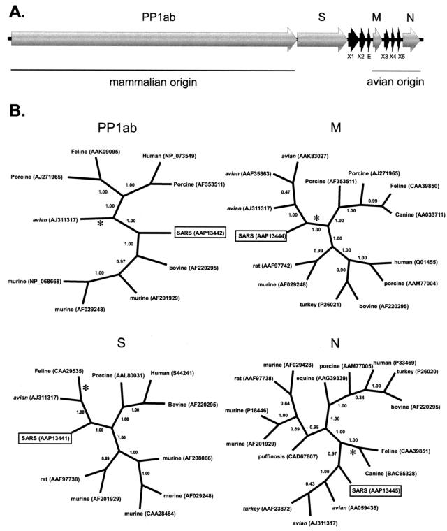
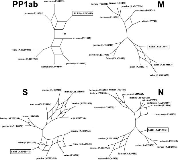
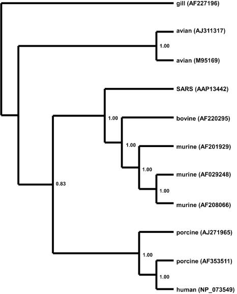
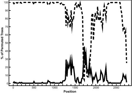
Severe acute respiratory syndrome (SARS) is a deadly form of pneumonia caused by a novel coronavirus, a viral family responsible for mild respiratory tract infections in a wide variety of animals including humans, pigs, cows, mice, cats, and birds. Analyses to date have been unable to identify the precise origin of the SARS coronavirus. We used Bayesian, neighbor-joining, and split decomposition phylogenetic techniques on the SARS virus replicase, surface spike, matrix, and nucleocapsid proteins to reveal the evolutionary origin of this recently emerging infectious agent. The analyses support a mammalian-like origin for the replicase protein, an avian-like origin for the matrix and nucleocapsid proteins, and a mammalian-avian mosaic origin for the host-determining spike protein. A bootscan recombination analysis of the spike gene revealed high nucleotide identity between the SARS virus and a feline infectious peritonitis virus throughout the gene, except for a 200- base-pair region of high identity to an avian sequence. These data support the phylogenetic analyses and suggest a possible past recombination event between mammalian-like and avian-like parent viruses. This event occurred near a region that has been implicated to be the human receptor binding site and may have been directly responsible for the switch of host of the SARS coronavirus from animals to humans.


Figures

FIG. 1.
FIG. 1.
(A) Organization of the SARS coronavirus genome. (B) Bayesian-inferred unrooted phylogenies of the SARS proteins, generated with MRBAYES version 3.04b using Jones-Taylor-Thornton protein weight matrix. Tree sampling was performed for 500,000 generations, and a majority-rule consensus tree was built with a burn-in of 100. The PP1ab phylogeny was based on a conserved 322-aa region, corresponding to aa 3230 to 3552 of the polyprotein. The S, M, and N genealogies are based on edited, whole-protein alignments of 840, 162, and 345 aa, respectively. Branch confidence values are given as PPs. Congruent trees were obtained by neighbor joining, with critical nodes being supported by bootstrap values that are >89%. Asterisks denote the location of midpoint rooting. Group 1 coronaviruses have initial capital letters. Group 2 coronaviruses are in lowercase letters. Group 3 coronaviruses are in italics.
FIG. 2.
FIG. 2.
Split decomposition graph of the SARS coronavirus genome. The PP1ab phylogeny was based on a conserved 322-aa region, corresponding to aa 3230 to 3552 of the polyprotein. The S, M, and N genealogies are based on edited, whole-protein alignments of 840, 162, and 345 aa, respectively. The asterisk on the PP1ab protein denotes the branch that is lost when the whole protein is analyzed.
FIG. 3.
FIG. 3.
Bayesian-inferred rooted phylogeny of the PP1ab polyprotein. Two conserved regions of the polyprotein, homologous to sequences found in the gill-associated okavirus, were concatenated and edited to eliminate gaps and ambiguously aligned regions. A phylogeny was generated on the resulting 880-aa alignment as described in the legend to Fig. 1. The tree was rooted on the gill-associated okavirus, a member of the Roniviridae, which is a sister family to the Coronaviridae. Branch confidence values are given as PPs.
FIG. 4.
FIG. 4.
Bootscan analysis of the S gene to detect recombination. Bootscanning was conducted with Simplot on a gapless nucleotide alignment, generated with ClustalX, with the SARS S gene as the query sequence. The dashed line denotes the feline infectious peritonitis virus (X06170), while the solid line denotes the avian infectious bronchitis virus (M95169). The greater the percentage of permuted trees, the greater the sequence identity to the SARS S sequence. Other taxa that were included in the alignment but had less than 10% permuted trees were murine hepatitis virus (AF208066) and human coronavirus OC43 (L14643).

References

    1. Alejska, M., A. Kurzynska-Kokorniak, M. Broda, R. Kierzek, and M. Figlerowicz. 2001. How RNA viruses exchange their genetic material. Acta Biochim. Pol. 48:391-407. - PubMed
    1. Anand, K., J. Ziebuhr, P. Wadhwani, J. R. Mesters, and R. Hilgenfeld. 2003. Coronavirus main proteinase (3CLpro) structure: basis for design of anti-SARS drugs. Science 300:1763-1767. - PubMed
    1. Bonavia, A., B. D. Zelus, D. E. Wentworth, P. J. Talbot, and K. V. Holmes. 2003. Identification of a receptor-binding domain of the spike glycoprotein of human coronavirus HCoV-229E. J. Virol. 77:2530-2538. - PMC - PubMed
    1. Chang, S. H., J. L. Bae, T. J. Kang, J. Kim, G. H. Chung, C. W. Lim, H. Laude, M. S. Yang, and Y. S. Jang. 2002. Identification of the epitope region capable of inducing neutralizing antibodies against the porcine epidemic diarrhea virus. Mol. Cell 14:295-299. - PubMed
    1. de Haan, C. A. M., H. Volders, C. A. Koetzner, P. S. Masters, and P. J. M. Rottier. 2002. Coronaviruses maintain viability despite dramatic rearrangements of the strictly conserved genome organization. J. Virol. 76:12491-12502. - PMC - PubMed

Publication types

MeSH terms

LinkOut - more resources