. 2004 Jan;78(1):197-205.
doi: 10.1128/jvi.78.1.197-205.2004.

Evasion of cellular antiviral responses by human cytomegalovirus TRS1 and IRS1

Affiliations

Evasion of cellular antiviral responses by human cytomegalovirus TRS1 and IRS1

Stephanie J Child et al. J Virol. 2004 Jan.

Abstract

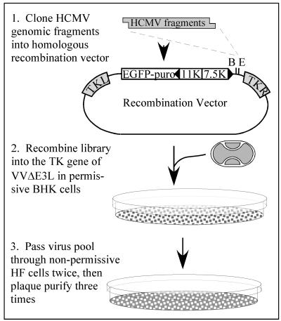
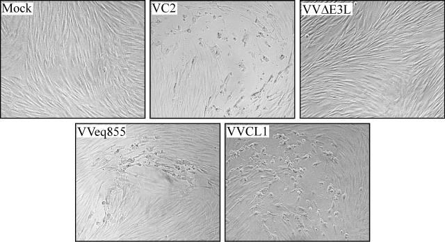
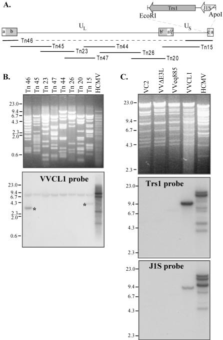
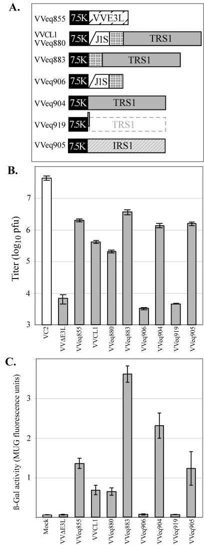
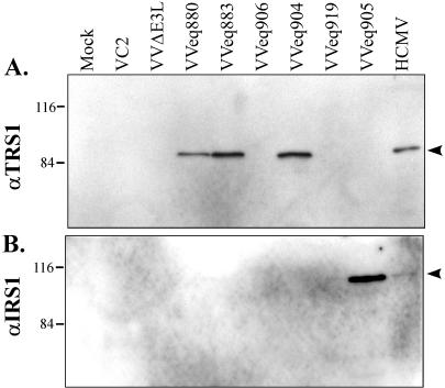
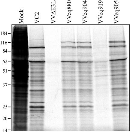
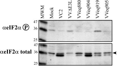
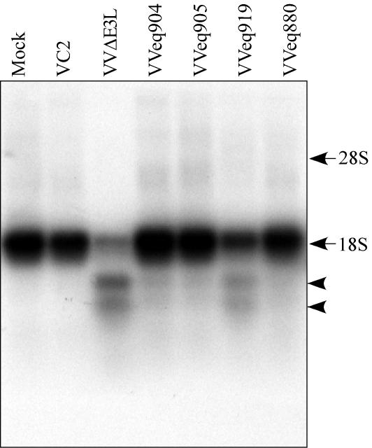
During infection with human cytomegalovirus (HCMV), cellular protein synthesis continues even as viral proteins are being synthesized in abundance. Thus, HCMV may have a mechanism for counteracting host cell antiviral pathways that act by shutting off translation. Consistent with this view, HCMV infection of human fibroblasts rescues the replication of a vaccinia virus mutant lacking the double-stranded RNA-binding protein gene E3L (VVdeltaE3L). HCMV also prevents the phosphorylation of the eukaryotic translation initiation factor eIF-2alpha, the activation of RNase L, and the shutoff of viral and cellular protein synthesis that otherwise result from VVdeltaE3L infection. To identify the HCMV gene(s) responsible for these effects, we prepared a library of VVdeltaE3L recombinants containing HCMV genomic fragments. By infecting nonpermissive cells with this library and screening for VV gene expression and replication, we isolated a virus containing a 2.8-kb HCMV fragment that rescues replication of VVdeltaE3L. The fragment comprises the 3' end of the J1S open reading frame through the entire TRS1 gene. Analyses of additional VVdeltaE3L recombinants revealed that the protein encoded by TRS1, pTRS1, as well as the closely related IRS1 gene, rescues VVdeltaE3L replication and prevent the shutoff of protein synthesis, the phosphorylation of eIF-2alpha, and activation of RNase L. These results demonstrate that TRS1 and IRS1 are able to counteract critical host cell antiviral response pathways.


Figures

FIG. 1.
FIG. 1.
Strategy for selection of HCMV fragments that rescue VVΔE3L replication. HCMV genomic DNA, digested with various enzyme combinations as described in Materials and Methods to yield fragments containing either BamHI (B)- or EcoRI (E)-compatible ends, was cloned into either of these sites in a VV homologous recombination vector. The insert is under the transcriptional control of the VV 7.5K promoter and, along with a VV11K promoter/EGFP-puromycin gene, is flanked by portions of the VV TK gene (TK left and TK right). The libraries of HCMV fragments were recombined into the TK locus of VVΔE3L in BHK cells, and the resulting virus pools were passed through VVΔE3L-nonpermissive HF in order to select recombinants containing HCMV genes that rescue VVΔE3L replication.
FIG. 2.
FIG. 2.
Plaque formation by VVCL1 in HF. HF were mock infected or infected with 10 to 100 PFU (as measured on BHK cells) of the indicated VV. Photomicrographs of plaques viewed 36 hpi by phase-contrast microscopy are shown. No plaques were detected in the mock-infected or VVΔE3L-infected wells (original magnification, ×200).
FIG. 3.
FIG. 3.
Southern blot analyses of VVCL1. (A) The HCMV genome contains unique long (UL) and short (US) segments flanked by inverted repeats (ab, b′a′c′, ca). The locations of the HCMV cosmid inserts and of the EcoRI/ApoI fragment present in VVCL1 are shown. (B) DNAs from HCMV and from HCMV cosmids were digested with EcoRI and XhoI, separated electrophoretically, stained with ethidium bromide (top), and then hybridized with 32P-labeled VVCL1 DNA. Autoradiography (bottom) identified specific bands in Tn46 and Tn15 (*). (C) DNA from HCMV or from the indicated VV was digested with HindIII and then viewed by ethidium bromide staining (top) and hybridized with TRS1 (middle) and J1S (bottom) probes. VCL1 and HCMV, but none of the other VVs, contained TRS1 and IRS1 sequences. Molecular size markers (in kilobases) are indicated.
FIG.4.
FIG.4.
Replication of VV in HF. (A) Recombinant VV viruses containing the 7.5K promoter driving expression of the E3L gene or of the indicated fragments from HCMV, including a portion of the J1S ORF, an intercistronic region (stippled), and the TRS1 or IRS1 ORF were constructed as described in Materials and Methods. VVeq880 contains the same HCMV insert as VVCL1, but they were made independently and used different recombination plasmids. Compared to VVeq904, VVeq919 contains a single-nucleotide insertion at the start of the TRS1 ORF. (B and C) HF were mock infected or infected with the indicated VV (MOI, 3). At 24 hpi (panel B), viral titers of freeze-thaw lysates were measured by plaque assays on BHK cells, and β-Gal activities (panel C) were measured as described in Materials and Methods. Means ± standard deviations of results for triplicate samples are shown.
FIG. 5.
FIG. 5.
Identification of protein products expressed by VVΔE3L recombinant viruses. BHK cells were infected with the different recombinant viruses as indicated in Materials and Methods. Cell lysates were prepared 24 h later and, along with lysates of HCMV-infected HF, were separated by SDS-PAGE and analyzed by immunoblot assay using antibody specific for the TRS1 protein (A) and the IRS1 protein (B). Molecular size markers (in kilodaltons) are shown to the left.
FIG. 6.
FIG. 6.
Protein synthesis in HF infected with various VVΔE3L recombinant viruses. HF were mock infected or infected with the indicated virus (MOI, 3) and labeled 24 h later for 1 h with [35S]methionine. Cell extracts were then prepared, and 20 μg of each was analyzed by SDS-PAGE and autoradiography. Molecular size markers (in kilodaltons) are indicated on the left.
FIG. 7.
FIG. 7.
Phosphorylation of eIF-2α after infection with the VVΔE3L recombinant viruses. HF were mock infected or infected with the indicated virus (MOI, 3). Cell lysates were prepared after 24 h, separated by SDS-PAGE, and analyzed by immunoblot assay using antibody specific for phosphorylated eIF-2α or total eIF-2α. Molecular size markers (in kilodaltons) are indicated to the left of each panel.
FIG. 8.
FIG. 8.
RNase L activity in cells infected with the VVΔE3L recombinant viruses. HF were mock infected or infected with the viruses indicated (MOI, 3). At 24 hpi, RNA was harvested, and 2 μg of each sample was analyzed by formaldehyde agarose gel electrophoresis and by Northern blot hybridization using a 32P-labeled 18S rRNA-specific probe. Characteristic 18S rRNA RNase L cleavage products (arrowheads) and the positions of intact rRNA bands made visible by ethidium bromide staining (arrows) are indicated.

References

    1. Abbate, J., J. C. Lacayo, M. Prichard, G. Pari, and M. A. McVoy. 2001. Bifunctional protein conferring enhanced green fluorescence and puromycin resistance. BioTechniques 31:336-340. - PubMed
    1. Beattie, E., K. L. Denzler, J. Tartaglia, M. E. Perkus, E. Paoletti, and B. L. Jacobs. 1995. Reversal of the interferon-sensitive phenotype of a vaccinia virus lacking E3L by expression of the reovirus S4 gene. J. Virol. 69:499-505. - PMC - PubMed
    1. Biegalke, B. J., and A. P. Geballe. 1990. Translational inhibition by cytomegalovirus transcript leaders. Virology 177:657-667. - PubMed
    1. Blankenship, C. A., and T. Shenk. 2002. Mutant human cytomegalovirus lacking the immediate-early TRS1 coding region exhibits a late defect. J. Virol. 76:12290-12299. - PMC - PubMed
    1. Brandt, T. A., and B. L. Jacobs. 2001. Both carboxy- and amino-terminal domains of the vaccinia virus interferon resistance gene, E3L, are required for pathogenesis in a mouse model. J. Virol. 75:850-856. - PMC - PubMed

Publication types

MeSH terms

LinkOut - more resources