. 2004 Jan;78(1):216-23.
doi: 10.1128/jvi.78.1.216-223.2004.

N-terminal domain of the murine coronavirus receptor CEACAM1 is responsible for fusogenic activation and conformational changes of the spike protein

Affiliations

N-terminal domain of the murine coronavirus receptor CEACAM1 is responsible for fusogenic activation and conformational changes of the spike protein

Hideka S Miura et al. J Virol. 2004 Jan.

Abstract

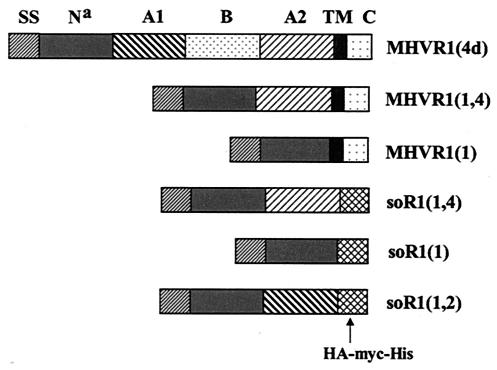
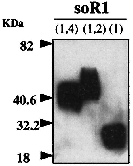
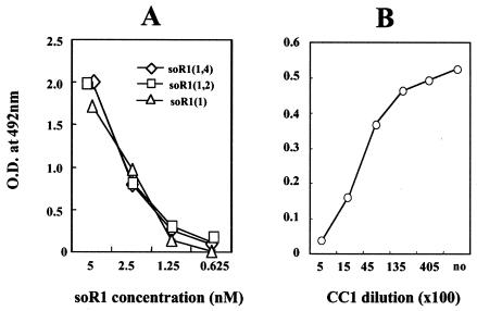
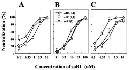
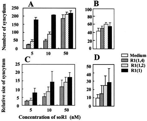
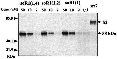
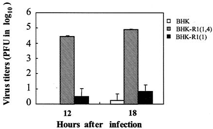
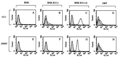
The mouse hepatitis virus (MHV) receptor (MHVR), CEACAM1, has two different functions for MHV entry into cells: binding to MHV spike protein (S protein) and activation of the S protein to execute virus-cell membrane fusion, the latter of which is accompanied by conformational changes of the S protein. The MHVR comprising the N-terminal and fourth domains [R1(1,4)] displays these two activities, and the N domain is thought to be critical for binding to MHV. In this study, we have addressed whether or not the N domain alone is sufficient for these activities. We examined three types of soluble form MHVR (soMHVR), one consisting of the N domain alone [soR1(1)], one with the N and second domains [soR1(1,2)], and one [soR1(1,4)] expressed by recombinant baculoviruses. We assessed the abilities of these three types of soMHVR to bind to MHV, activate fusogenicity, and induce conformational changes of the S protein. All three types of soMHVR similarly bound to MHV, as examined by a solid-phase binding assay and neutralized MHV infectivity. They also activated S protein fusogenicity and induced its conformational changes with similar levels of efficiency. However, R1(1) expressed on the BHK cell surface failed to serve as a receptor in spite of a sufficient level of expression. The inability of expressed R1(1) to work as a receptor was due to the inaccessibility of virions to R1(1); however, these were accessible using the MHVR-specific monoclonal antibody CC1. These results collectively indicated that the N domain retains all biological activities necessary for receptor function.


Figures

FIG. 1.
FIG. 1.
Schematic diagram of the CEACAM1 used in the present study. Prototype CEACAM1 (MHVR) is composed of four ectodomains linked with TM and Cy, and genetically manipulated MHVRs lack some of those domains and regions. Three different soluble forms have three tags (HA, myc, and 6×His) that replace TM and Cy, and the other two MHVRs used for expression on cell membranes lack one or two ectodomains.
FIG. 2.
FIG. 2.
Western blot analysis of the purified soMHVR. These soR1s were expressed by recombinant baculoviruses and purified by affinity chromatography using 6×His. After electrophoresis in an SDS-10% polyacrylamide gel, proteins were blotted onto nitrocellulose membrane and soR1s were detected with anti-HA MAb and anti-mouse IgG labeled with HRPO.
FIG. 3.
FIG. 3.
Solid-phase virus-binding assay. (A) Various soR1s were prepared on 96-well plates by incubating at 4°C overnight or 37°C for 2 h. JHMV was added and incubated at 37°C for 1 h, and its binding was monitored using anti-JHMV S MAb and anti-mouse IgG labeled with HRPO. (B) To determine the levels of binding specificity, soR1(1,4) prepared in 96-well plates was treated with CC1 at 37°C for 1 h and then incubated with JHMV. JHMV binding to soR1(1,4) was monitored as described above. Results are representative of multiple independent experiments.
FIG. 4.
FIG. 4.
Virus neutralization by soR1s. wt JHMV cl-2 (A), srr7 (B), or MHV-A59 (C) (about 100 PFU/20 μl) was mixed with an equal volume of soR1s and incubated at RT (A) or 37°C (B and C) for 50 min, and the residual infectivity was measured on DBT cells. Inhibition of fusion was estimated by comparing the infectivity after incubation in the absence of soR1s. Error bars represent standard deviations of the results for three independent samples.
FIG. 5.
FIG. 5.
Fusogenic activation of JHMV by soR1s. srr7 (A and C)- or wt JHM (B and D)-infected DBT cells (104) were overlaid onto BHK cell monolayers prepared in 24-well plates and incubated for 15 h in the presence or absence of soR1. The numbers (A and B) or sizes (C and D) of syncytia were measured after staining with crystal violet. Various soR1s were employed for the wt (B and D) at a concentration of 10 nM. Error bars represent standard deviations of the results for three independent samples.
FIG. 6.
FIG. 6.
Conformational changes of the S2 by soR1s. srr7 was mixed with soR1s or left untreated (−), incubated at 37°C for 30 min, and subsequently digested with proteinase K (20 mg/ml) at 4°C for 20 min. Following SDS-PAGE and blotting onto the nitrocellulose membrane, proteinase-resistant fractions of 58 kDa were detected using anti-S2 MAb 10G. Lane srr7, S protein not treated by soR1 and proteinase K.
FIG. 7.
FIG. 7.
Virus growth in BHK cells expressing various R1s. BHK cells expressing R1(1,4), R1(1), or no R1 and designated BHK-R1(1,4), BHK-R1(1), or BHK cells, respectively, were infected with wt JHMV cl-2 at an MOI of 0.1 and incubated for 12 or 18 h. Virus titers in the culture were measured using DBT cells. Bars show the standard deviations of the results for three independent samples.
FIG. 8.
FIG. 8.
Flow cytometric analysis of MHVRs expressed on cell membrane, as examined using MAb CC1 (A to D), and of JHMV that bound to those MHVRs (E to H). (A to D) Cells expressing various MHVRs were dispersed by proteinase and reacted with anti-MHVR MAb CC1 (open histograms) at 4°C for 30 min and subsequently with anti-mouse IgG labeled with Alexa Fluor 488 at 4°C for 30 min. Mouse IgG1 was used as an isotype control (shaded histograms). After fixation in 1% paraformaldehyde, cells were analyzed on a FACSCalibur apparatus using CellQuest software. (E to H) Cells (dispersed as described above) were mixed with 2 × 10⁵ PFU of JHMV (open histograms) and incubated at 4°C for 30 min. Then, the cells were reacted with anti-MHV S protein MAb no. 2 or 7 at 4°C for 30 min and subsequently with the above-described anti-mouse IgG. Finally, the cells were analyzed as described above. IgG2a was used as an isotype control (shaded histograms). FL1-H, log intensity of fluorescence.

Similar articles

Cited by

References

    1. Beauchemin, N., et al. 1999. Redefined nomenclature for members of the carcinoembryonic antigen family. Exp. Cell Res. 252:243-249. - PubMed
    1. Chen, D. C., and P. S. Kim. 1998. HIV entry and its inhibition. Cell 93:681-684. - PubMed
    1. Dalgleish, A. G., P. C. Beverley, P. R. Clapham, D. H. Crawford, M. F. Greaves, and R. A. Weiss. 1984. The CD4 (T4) antigen is an essential component of the receptor for the AIDS retrovirus. Nature 312:763-766. - PubMed
    1. Drosten, C., et al. 2003. Identification of a novel coronavirus in patients with severe acute respiratory syndrome. N. Engl. J. Med. 348:1967-1976. - PubMed
    1. Dveksler, G. S., A. A. Basile, C. B. Cardellichio, and K. V. Holmes. 1995. Mouse hepatitis virus receptor activities of an MHVR/mph chimera and MHVR mutants lacking N-linked glycosylation of the N-terminal domain. J. Virol. 69:543-546. - PMC - PubMed

Publication types

MeSH terms

LinkOut - more resources