. 2004 Jan;78(1):224-39.
doi: 10.1128/jvi.78.1.224-239.2004.

Herpes simplex virus type 1 immediate-early gene expression is required for the induction of apoptosis in human epithelial HEp-2 cells

Affiliations

Herpes simplex virus type 1 immediate-early gene expression is required for the induction of apoptosis in human epithelial HEp-2 cells

Christine M Sanfilippo et al. J Virol. 2004 Jan.

Abstract

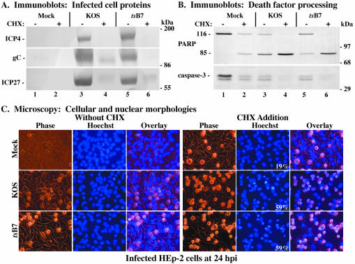
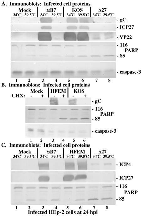
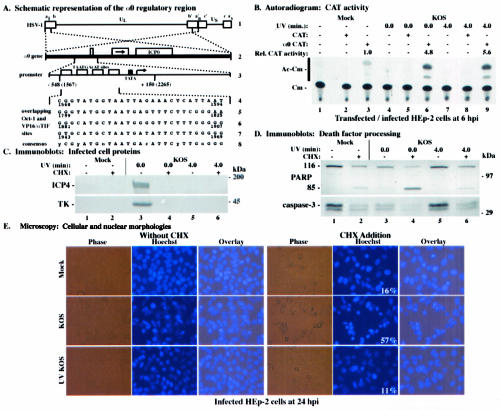
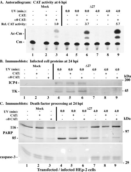
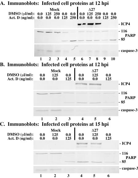
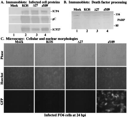
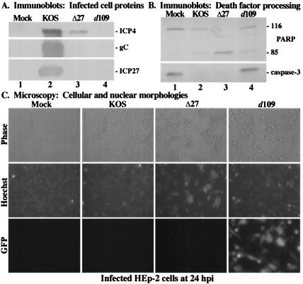
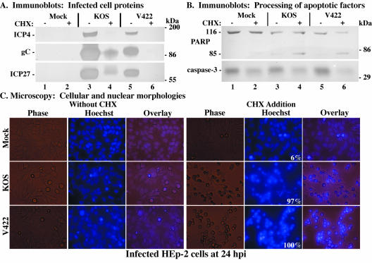
Wild-type herpes simplex virus type 1 (HSV-1) induces apoptosis in human epithelial HEp-2 cells, but infected cell proteins produced later in infection block the process from killing the cells. Thus, HSV-1 infection in the presence of the translational inhibitor cycloheximide (CHX) results in apoptosis. Our specific goal was to gain insight as to the viral feature(s) responsible for triggering apoptosis during HSV-1 infection. We now report the following. (i) No viral protein synthesis or death factor processing was detected after infection with HSV-1(HFEMtsB7) at 39.5 degrees C; this mutant virus does not inject its virion DNA into the nucleus at this nonpermissive temperature. (ii) No death factor processing or apoptotic morphological changes were detected following infection with UV-irradiated, replication-defective viruses possessing transcriptionally active incoming VP16. (iii) Addition of the transcriptional inhibitor actinomycin D prevented death factor processing upon infection with the apoptotic, ICP27-deletion virus HSV-1(vBSDelta27). (iv) Apoptotic morphologies and death factor processing were not observed following infection with HSV-1(d109), a green fluorescent protein-expressing recombinant virus possessing deletions of all five immediate-early (IE) (or alpha) genes. (v) Finally, complete death factor processing was observed upon infection with the VP16 transactivation domain-mutant HSV-1(V422) in the presence of CHX. Based on these findings, we conclude that (vi) the expression of HSV-1 alpha/IE genes is required for the viral induction of apoptosis and (vii) the transactivation activity of VP16 is not necessary for this induction.


Figures

FIG. 1.
FIG. 1.
Immune reactivities of infected cell proteins (A) and death factors (B) and cellular and nuclear morphologies (C) in infected HEp-2 cells. Whole-cell extracts were prepared at 24 hpi from mock-infected cells or cells infected with HSV-1(KOS1.1) or HSV-1(HFEMtsB7) in the absence (−) or presence (+) of CHX, separated in a denaturing gel, transferred to nitrocellulose, and probed with anti-ICP4, -gC, and -ICP27 (A) or -PARP and -caspase-3 (B) antibodies as described in Materials and Methods. Full-length (116,000-molecular-weight) (116) PARP and processed (85,000-molecular-weight) (85) PARP are shown. The locations of prestained molecular weight markers are indicated in the right margin. Phase-contrast, corresponding fluorescence (Hoechst), and merged (Overlay) images were obtained for mock-, HSV-1(KOS1.1)-, and HSV-1(HFEMtsB7)-infected HEp-2 cells in the absence (Without CHX) and presence (CHX Addition) of CHX; stained with Hoechst H33258 DNA dye; and visualized at 24 hpi as described in Materials and Methods. These cells were used to prepare the whole-cell extracts shown in panels A and B. White numbers in panels refer to the percentages of cells showing apoptotic condensed chromatin. Magnification, ×40.
FIG. 2.
FIG. 2.
Immune reactivities of infected cell proteins and death factors in infected HEp-2 cells. (A) Whole-cell extracts were prepared at 24 hpi from mock-infected cells or cells infected with HSV-1(HFEMtsB7), HSV-1(KOS1.1), or HSV-1(vBSΔ27) (Δ27) at the permissive (34°C) and nonpermissive (39.5°C) temperatures as described in Materials and Methods. Immunoblot analyses utilized anti-gC, -ICP27, -VP22, -PARP, and -caspase-3 antibodies. (B) Whole-cell extracts were prepared 24 hpi from mock-infected cells or cells infected with HSV-1(HFEM) or HSV-1(KOS1.1) in the absence (−) or presence (+) of CHX. Immunoblot analyses utilized anti-gC, -PARP, and -caspase-3 antibodies. (C) Whole-cell extracts were prepared at 24 hpi from mock-infected cells or cells infected with HSV-1(HFEMtsB7), HSV-1(HFEM), or HSV-1(vBSΔ27) at the permissive (34°C) and nonpermissive (39.5°C) temperatures. Immunoblot analyses utilized anti-ICP4, -ICP27, or -PARP antibodies. 116 and 85, full-length and processed PARP, respectively.
FIG. 3.
FIG. 3.
(A) Schematic diagram of the HSV-1 α0 promoter region contained in the α0 CAT plasmid. (Line 1) HSV-1 DNA genome, indicating the unique sequences (thin lines) flanked by the inverted repeats (boxes). The letters above line 1 designate the a sequence (al) and b sequence (b) of the long component, the unique sequence of the long component (UL), the repetitions of the b and of a variable (n) number of a sequences (an), the inverted c sequence, the unique sequence of the short component (US), and finally the terminal a sequence (as) of the short component (41, 42, 57). (Line 2) α0 gene, present in two copies (dotted lines) in the UL inverted repeats of the HSV-1 DNA (thick line). The three exons encoding the ICP0 protein are indicated by boxes, separated by introns (thin lines). The arrow in line 2 indicates the direction of protein translation. (Line 3) α0 promoter region (dotted lines). Open boxes indicate the four TAATGArAT sequences, which are the cis sites for the induction of α genes by αTIF (18, 38, 39). The cellular TATA-binding protein (TBP) binding site (TATA sequence) is designated by a filled box. The arrow indicates the transcription start site. Vertical lines below line 3 show the section of the α0 promoter region contained in the α0 CAT plasmid. Nucleotide positions are shown with reference to both the transcriptional start site and coordinates in the HSV-1 genome (in parentheses) (41, 42). (Lines 4 to 7) Nucleotide sequences of the overlapping Oct-1 and VP16/αTIF binding sites. The numbers below each line give the genome positions of each TAATGArAT sequence. (Line 8) Consensus TAATGArAT sequence. y is pyrimidine, r is purine, and n is any nucleotide. (B) Autoradiographic image depicting CAT activity in HEp-2 cells infected with HSV-1(KOS1.1). HEp-2 cells were transfected with 2.5 μg of either CAT or α0 CAT plasmid and infected 48 h posttransfection with untreated or UV-treated HSV-1(KOS1.1). Cytoplasmic extracts were prepared at 6 hpi and assayed for CAT activity as described in Materials and Methods. Relative CAT activity refers to the level of increase in CAT activity compared to that in mock-infected α0 CAT-transfected cells. Unmodified (Cm) and acetylated (Ac-Cm) forms of chloramphenicol are indicated on the left. (C and D) Immune reactivities of infected cell proteins (C) and death factors (D) in infected HEp-2 cells. Whole-cell extracts prepared at 24 hpi from mock-infected cells or cells infected with untreated or UV-treated HSV-1(KOS1.1) in the absence (−) or presence (+) of the protein synthesis inhibitor CHX (10 mg/ml) were separated in a denaturing gel, transferred to nitrocellulose, and probed with the anti-ICP4 or -TK (C) or -PARP or -caspase-3 (D) antibodies, as described in Materials and Methods. UV exposure times refer to untreated and UV-treated viruses, respectively. 116 and 85, full-length and processed PARP, respectively. The locations of prestained molecular weight markers are indicated in the right margins. (E) Phase-contrast, corresponding fluorescence (Hoechst), and merged (Overlay) visualizations of infected HEp-2 cells. HEp-2 cells at 24 h after mock infection or infection with untreated or UV-treated HSV-1(KOS1.1) in the absence (Without CHX) or presence (CHX Addition) of CHX were visualized as described in Materials and Methods. Numbers in panels (white) refer to the percentages of cells showing apoptotic condensed chromatin. Magnification, ×40. These cells were used to prepare the whole-cell extracts shown in panels C and D.
FIG. 4.
FIG. 4.
Autoradiographic images (A) and immune reactivities (B and C) of HEp-2 cells infected with untreated or UV-treated HSV-1(vBSΔ27) (Δ27). HEp-2 cells were transfected with 2.5 μg of either CAT or α0 CAT plasmid and infected 48 h posttransfection with untreated or UV-treated HSV-1(vBSΔ27). (A) CAT activity from infected HEp-2 cell cytoplasmic extracts prepared at 6 hpi. Relative CAT activity refers to the increase (n-fold) in CAT activity compared to that of mock-infected α0 CAT-transfected cells. Unmodified and acetylated forms of chloramphenicol are indicated in the left margin. Whole-cell extracts prepared from infected HEp-2 cells at 24 hpi were separated in a denaturing gel, transferred to nitrocellulose, and probed with the anti-ICP4 and -TK (B) or -PARP and -caspase-3 (C) antibodies. UV exposure times refer to untreated and UV-treated viruses, respectively. The locations of prestained molecular mass markers are indicated in the right margins. 116 and 85, full-length and processed PARP, respectively.
FIG. 5.
FIG. 5.
Immune reactivities of infected cell proteins and death factors at 12 hpi (A and B) or 15 hpi (C) from vBSΔ27-infected HEp-2 cells plus actinomycin D. Whole-cell extracts were prepared from mock-infected cells or cells infected with HSV-1(vBSΔ27) (Δ27) in the absence or presence of the transcription inhibitor actinomycin D or DMSO solvent as described in Materials and Methods. Immunoblot analyses utilized anti-ICP4, -PARP, and -caspase-3 antibodies.
FIG. 6.
FIG. 6.
Immune reactivities of infected cell proteins (A) and death factors (B) and cell morphologies (C) of infected FO6 cells. Whole-cell extracts were prepared at 24 hpi from mock-infected cells or cells infected with HSV-1(KOS1.1), HSV-1(vBSΔ27) (Δ27), or HSV-1(d109) as described in Materials and Methods. Immunoblot analyses utilized anti-ICP4, -gC, and -ICP27 antibodies (A) or -PARP antibodies (B). 116 and 85, full-length and processed PARP, respectively. (C) Phase-contrast and fluorescence (Hoechst and GFP) images of corresponding infected FO6 cells. Mock-, HSV-1(KOS1.1)-, HSV-1(vBSΔ27)-, and HSV-1(d109)-infected FO6 cells were visualized at 24 hpi as described in Materials and Methods. Magnification, ×40. These cells were used to prepare the whole-cell extracts shown in panels A and B.
FIG. 7.
FIG. 7.
Immune reactivities of infected cell proteins (A) and death factors (B) and cell morphologies (C) of infected HEp-2 cells. Whole-cell extracts were prepared at 24 hpi from mock-infected cells or cells infected with HSV-1(KOS1.1), HSV-1(vBSΔ27) (Δ27), or HSV-1(d109) as described in Materials and Methods. Immunoblot analyses utilized anti-ICP4, -gC, and -ICP27 antibodies (A) or -PARP and -caspase-3 antibodies (B). 116 and 85, full-length and processed PARP, respectively. (C) Phase-contrast and fluorescence (Hoechst and GFP) images of corresponding infected HEp-2 cells. Mock-, HSV-1(KOS1.1)-, HSV-1(vBSΔ27)-, and HSV-1(d109)-infected HEp-2 cells were visualized at 24 hpi as described in Materials and Methods. Magnification, ×40. These cells were used to prepare the whole-cell extracts shown in panels A and B.
FIG. 8.
FIG. 8.
Immune reactivities of infected cell proteins (A) and death factors (B) and cell morphologies (C) of infected HEp-2 cells. Whole-cell extracts were prepared at 24 hpi from mock-infected cells or cells infected with HSV-1(KOS1.1) or HSV-1(V422) in the absence (−) or presence (+) of CHX as described in Materials and Methods. Immunoblot analyses utilized anti-ICP4, -gC, and -ICP27 antibodies (A) or -PARP and -caspase-3 antibodies (B). The locations of prestained molecular mass markers are indicated in the right margins. 116 and 85, full-length and processed PARP, respectively. (C) Phase-contrast, fluorescence (Hoechst), and merged (Overlay) images of corresponding infected HEp-2 cells. Mock-, HSV-1(KOS1.1)-, and HSV-1(V422)-infected HEp-2 cells in the absence (Without CHX) and presence (CHX Addition) of CHX were visualized at 24 hpi as described in Materials and Methods. Numbers in panels (white) refer to the percentage of cells showing apoptotic condensed chromatin. Magnification, ×40. These cells were used to prepare the whole-cell extracts shown in panels A and B.

Similar articles

Cited by

References

    1. Aubert, M., and J. A. Blaho. 1999. The herpes simplex virus type 1 regulatory protein ICP27 is required for the prevention of apoptosis in infected human cells. J. Virol. 73:2803-2813. - PMC - PubMed
    1. Aubert, M., and J. A. Blaho. 2001. Modulation of apoptosis during herpes simplex virus infection in human cells. Microbes Infect. 3:859-866. - PubMed
    1. Aubert, M., J. O'Toole, and J. A. Blaho. 1999. Induction and prevention of apoptosis in human HEp-2 cells by herpes simplex virus type 1. J. Virol. 73:10359-10370. - PMC - PubMed
    1. Aubert, M., S. A. Rice, and J. A. Blaho. 2001. Accumulation of herpes simplex virus type 1 early and leaky-late proteins correlates with apoptosis prevention in infected human HEp-2 cells. J. Virol. 75:1013-1030. - PMC - PubMed
    1. Avitabile, E., S. Di Gaeta, M. R. Torrisi, P. L. Ward, B. Roizman, and G. Campadelli-Fiume. 1995. Redistribution of microtubules and Golgi apparatus in herpes simplex virus-infected cells and their role in viral exocytosis. J. Virol. 69:7472-7482. - PMC - PubMed

Publication types

MeSH terms

LinkOut - more resources