. 2004 Jan;78(1):257-65.
doi: 10.1128/jvi.78.1.257-265.2004.

Evidence for an essential catalytic role of the F10 protein kinase in vaccinia virus morphogenesis

Affiliations

Evidence for an essential catalytic role of the F10 protein kinase in vaccinia virus morphogenesis

Patricia Szajner et al. J Virol. 2004 Jan.

Abstract

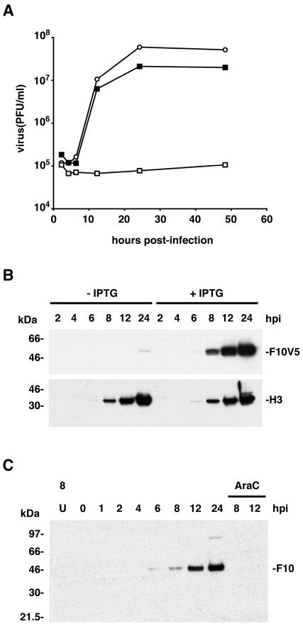
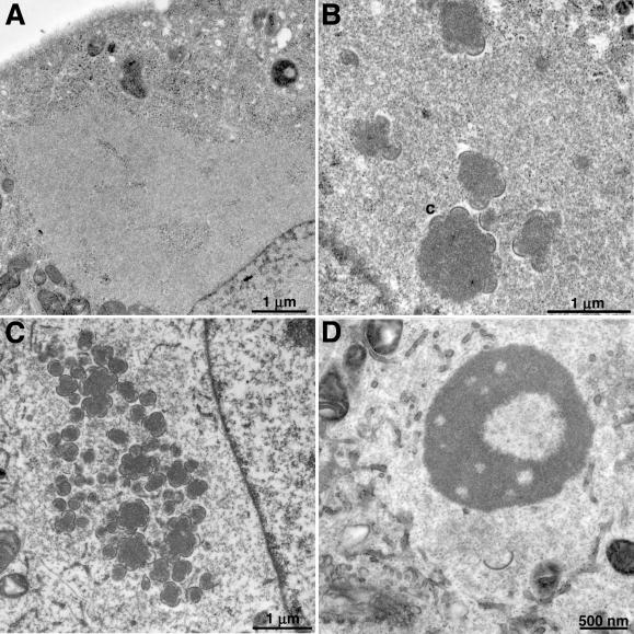
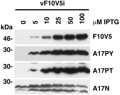
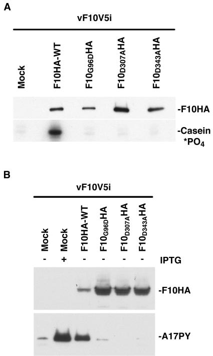
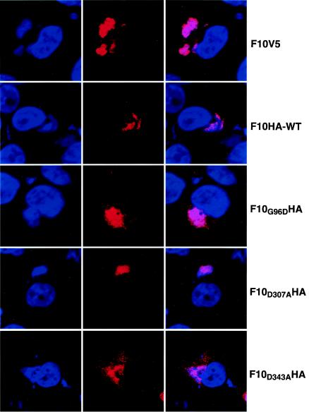
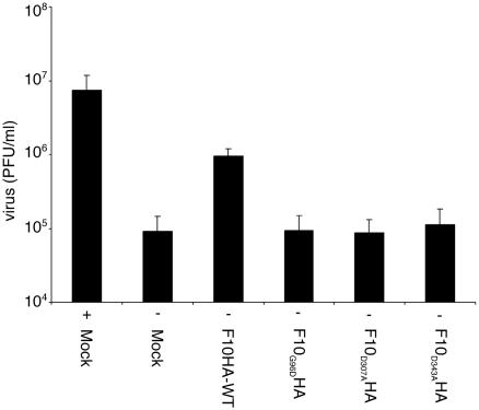
Temperature-sensitive mutants of vaccinia virus, with genetic changes that map to the open reading frame encoding the F10 protein kinase, exhibit a defect at an early stage of viral morphogenesis. To further study the role of the enzyme, we constructed recombinant vaccinia virus vF10V5i, which expresses inducible V5 epitope-tagged F10 and is dependent on a chemical inducer for plaque formation and replication. In the absence of inducer, viral membrane formation was delayed and crescents and occasional immature forms were detected only late in infection. When the temperature was raised from 37 to 39 degrees C, the block in membrane formation persisted throughout the infection. The increased stringency may be explained by a mild temperature sensitivity of the wild-type F10 kinase, which reduced the activity of the very small amount expressed in the absence of inducer, or by the thermolability of an unphosphorylated kinase substrate or uncomplexed F10-interacting protein. Further analyses demonstrated that tyrosine and threonine phosphorylation of the A17 membrane component was inhibited in the absence of inducer. The phosphorylation defect could be overcome by transfection of plasmids that express wild-type F10, but not by plasmids that express F10 with single amino acid substitutions that abolished catalytic activity. Although the mutated forms of F10 were stable and concentrated in viral factories, only the wild-type protein complemented the assembly and replication defects of vF10V5i in the absence of inducer. These studies provide evidence for an essential catalytic role of the F10 kinase in vaccinia virus morphogenesis.


Figures

FIG. 1.
FIG. 1.
Effects of IPTG on the replication of vF10V5i and synthesis of the F10 protein. (A) Effect of IPTG on virus yield. BS-C-1 cells were infected with VV WR (○) or vF10V5i in the absence (□) or presence (▪) of 50 μM IPTG. Cells were harvested at the indicated times after infection and virus titers were determined by plaque assay in the presence of IPTG. (B) Effect of IPTG on F10V5 and H3 protein synthesis. BS-C-1 cells were infected at a multiplicity of infection of 10 with vF10V5i in the presence or absence of 50 μM IPTG and harvested at intervals between 2 and 24 h postinfection (hpi). Proteins from total cell lysates were resolved by electrophoresis on a 10 to 20% polyacrylamide gradient gel in SDS-Tricine buffer, analyzed by Western blotting using anti-V5 or anti-H3 antibody, and detected by chemiluminescence. The numbers on the left correspond to the molecular masses of marker proteins. The positions of the F10V5 and H3 bands are indicated on the right. (C) Temporal synthesis of F10 in cells infected with WR. BS-C-1 cells were mock infected for 8 h (U) or infected at a multiplicity of infection of 10 in the absence or presence of AraC and harvested between 0 and 24 h postinfection (hpi). Proteins from whole-cell extracts were analyzed by Western blotting as in panel A except that the antiserum was prepared against a peptide composed of amino acids 5 to 20 of F10.
FIG. 2.
FIG. 2.
Electron microscopy of cells infected with vF10V5i. BS-C-1 cells were infected with vF10V5i at a multiplicity of infection of 10 at 37°C (A, B, C) or 39°C (D) in the absence of IPTG. Eight (A), 12 (B), or 24 (C and D) h after infection, the cells were fixed and prepared for transmission electron microscopy essentially as described previously (20). Electron micrographs are shown, with the scales indicated by bars. Abbreviations: C, crescents; nu, nucleoid within an IV.
FIG. 3.
FIG. 3.
Effect of IPTG on the synthesis of F10V5 and on the posttranslational modification of A17. BS-C-1 cells were infected with vF10V5i in the presence of 0 to 100 μM IPTG. Twenty-four hours after infection, the cells were harvested and the proteins were analyzed by electrophoresis on a 4 to 20% polyacrylamide gradient gel in SDS-Tris-glycine buffer. The proteins were then transferred to a nitrocellulose membrane and incubated with the anti-V5 (F10V5), anti-pTyr (A17PY), anti-pThr (A17PT), or anti-A17N antibody. The bands were detected by chemiluminescence. The numbers on the left correspond to the molecular masses of the marker proteins.
FIG. 4.
FIG. 4.
In vitro and in vivo activity of mutated F10 kinase. (A) In vitro activity. BS-C-1 cells were infected with vF10V5i in the absence of IPTG and either mock transfected or transfected with expression plasmids encoding HA epitope-tagged wild-type F10 (F10HA-WT) or mutated F10 (F10G96DHA, F10D307AHA, and F10D343AHA). Twenty-four hours after infection, the cells were harvested and the wild-type and mutated forms of F10 were bound to an anti-HA affinity matrix. The bound proteins were analyzed by electrophoresis on a 4 to 20% polyacrylamide gradient gel in SDS-Tris-glycine buffer followed by Western blotting using an anti-HA antibody conjugated to horseradish peroxidase. Kinase assays were performed in kinase buffer, with F10HA proteins still attached to the affinity matrix, 20 μM [γ-32P]ATP, and 1 μg of casein. After incubation for 10 min at 30°C, samples were denatured with SDS and analyzed on a 4 to 20% polyacrylamide gradient gel in SDS-Tris-glycine buffer. Proteins were visualized by autoradiography. (B) BS-C-1 cells were infected with vF10V5i in the presence of 50 μM IPTG or in the absence of IPTG and transfected as for panel A. Twenty-four hours after infection, the cells were harvested and total cell lysates were prepared. The proteins were analyzed by electrophoresis on a 10 to 20% polyacrylamide gradient gel in SDS-Tricine buffer. The proteins were then transferred to a nitrocellulose membrane and incubated with anti-HA and anti-pTyr antibodies to detect F10 and tyrosine-phosphorylated A17, respectively. The bands were detected by chemiluminescence.
FIG. 5.
FIG. 5.
Localization of F10 by confocal microscopy. HeLa cells were infected with vF10V5i at a multiplicity of infection of 5 in the absence or presence of 25 μM IPTG and transfected with plasmids as described in the legend for Fig. 4. Eight hours after infection, the cells were fixed with 4% paraformaldehyde in PBS for 20 min, washed and permeabilized with 0.1% Triton X-100 in PBS for 7 min, and blocked with 1% bovine serum albumin in PBS for 10 min. The cells infected in the presence of IPTG were stained with the anti-V5 MAb to detect the induced F10V5 protein, while those infected in the absence of the inducer and transfected with plasmids were stained with an anti-HA MAb (column 2). In each case, rhodamine red X-conjugated goat anti-mouse antibody was used as the secondary antibody. DNA in nuclei and viral factories was stained with DAPI (column 1). The cells were examined by confocal microscopy and merged images are shown in column 3. Colors: red, anti-V5 or anti-HA antibody; blue, DAPI.
FIG. 6.
FIG. 6.
Transcomplementation of virus infectivity. HeLa cells were infected and transfected as described in the legend for Fig. 4 with (+) or without (−) 50 μM IPTG. After 24 h, the cells were harvested and the virus titers were determined by plaque assay in the presence of 50 μM IPTG. Values are the averages plus standard deviations (error bars) of three independent transfection experiments.

References

    1. Betakova, T., E. J. Wolffe, and B. Moss. 1999. Regulation of vaccinia virus morphogenesis: phosphorylation of the A14L and A17L membrane proteins and C-terminal truncation of the A17L protein are dependent on the F10L protein kinase. J. Virol. 73:3534-3543. - PMC - PubMed
    1. Chiu, W. L., and W. Chang. 2002. Vaccinia virus J1R protein: a viral membrane protein that is essential for virion morphogenesis. J. Virol. 76:9575-9587. - PMC - PubMed
    1. da Fonseca, F. G., A. Weisberg, E. J. Wolffe, and B. Moss. 2000. Characterization of the vaccinia virus H3L envelope protein: topology and posttranslational membrane insertion via the C-terminal hydrophobic tail. J. Virol. 74:7508-7517. - PMC - PubMed
    1. Derrien, M., A. Punjabi, R. Khanna, O. Grubisha, and P. Traktman. 1999. Tyrosine phosphorylation of A17 during vaccinia virus infection: involvement of the H1 phosphatase and the F10 kinase. J. Virol. 73:7287-7296. - PMC - PubMed
    1. De Silva, F. S., and B. Moss. 2003. Vaccinia virus uracil DNA glycosylase has an essential role in DNA synthesis that is independent of its glycosylase activity: catalytic site mutations reduce virulence but not virus replication in cultured cells. J. Virol. 77:159-166. - PMC - PubMed

Substances

LinkOut - more resources