. 2004 Jan;78(1):266-74.
doi: 10.1128/jvi.78.1.266-274.2004.

Physical and functional interactions between vaccinia virus F10 protein kinase and virion assembly proteins A30 and G7

Affiliations

Physical and functional interactions between vaccinia virus F10 protein kinase and virion assembly proteins A30 and G7

Patricia Szajner et al. J Virol. 2004 Jan.

Abstract

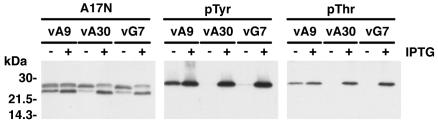
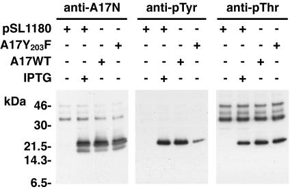
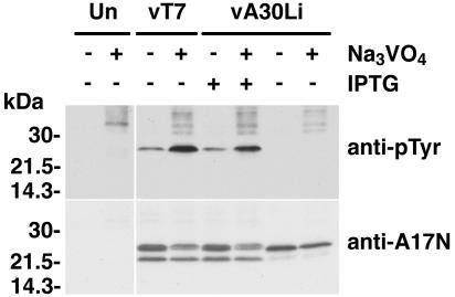
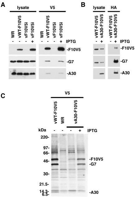
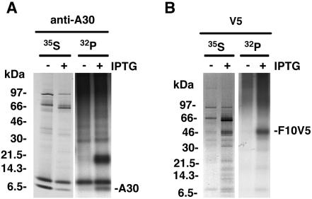
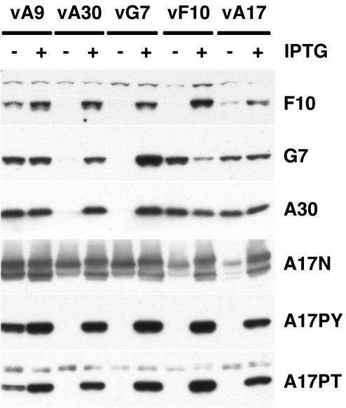
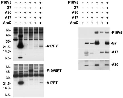
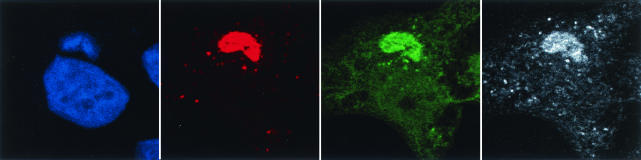
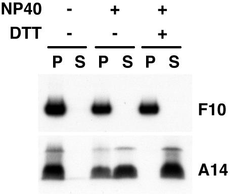
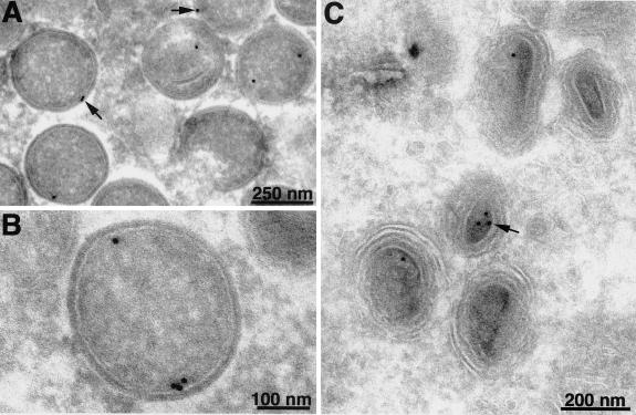
An early step in vaccinia virus morphogenesis, the association of crescent membranes with electron-dense granular material, is perturbed when expression of the viral protein encoded by the A30L or G7L open reading frame is repressed. Under these conditions, we found that phosphorylation of the A17 membrane protein, which is mediated by the F10 kinase, was severely reduced. Furthermore, A30 and G7 stimulated F10-dependent phosphorylation of A17 in the absence of other viral late proteins. Evidence for physical interactions between A30, G7, and F10 was obtained by their coimmunoprecipitation with antibody against A30 or F10. In addition, phosphorylation of A30 was dependent on the F10 kinase and autophosphorylation of F10 was stimulated by A30 and G7. Nevertheless, the association of A30, G7, and F10 occurred even with mutated, catalytically inactive forms of F10. Just as A30 and G7 are mutually dependent on each other for stability, F10 was nearly undetectable in the absence of A30 and G7. The reverse is not true, however, as repression of F10 did not diminish A30 or G7. Interaction of F10 with A30 and G7 presumably occurred within the virus factory areas of the cytoplasm, where each was concentrated. F10 localized predominantly in the cortical region of immature virions, beneath the membrane where A17 is located. F10 remained associated with the particulate core fraction of mature virions after treatment with a nonionic detergent and reducing agent. The formation of protein complexes such as the one involving A30, G7, and F10 may be a mechanism for the regulated packaging and processing of virion components.


Figures

FIG. 1.
FIG. 1.
A17 is not phosphorylated on tyrosine and threonine residues in the absence of A30 and G7 expression. (A) BS-C-1 cells were infected with vA9i (vA9), vA30Li (vA30), or vG7Li (vG7) virus in the presence (+) or absence (−) of 50 μM IPTG, as indicated. Twenty-four hours after infection, cells were harvested and total cell lysates were analyzed by electrophoresis in an SDS-4 to 20% gradient polyacrylamide gel in Tris-glycine buffer followed by Western blotting using the anti-A17N, anti-pTyr, and anti-pThr antisera. The migration positions and masses of marker proteins are indicated on the left.
FIG. 2.
FIG. 2.
Tyrosine and threonine phosphorylation of A17. BS-C-1 cells were infected with vA17Li at a multiplicity of infection of 5 PFU per cell in the presence (+) or absence (−) of 50 μM IPTG. Infected cells were transfected (+) with different combinations of plasmids containing a wild-type (A17WT) or mutated (A17Y203F) form of the A17L ORF under the control of the P11 promoter or with vector alone (pSL1180), as indicated. At 24 h, cells were harvested and total cell lysates were analyzed by electrophoresis in an SDS-10 to 20% polyacrylamide gel in Tricine buffer followed by Western blotting using anti-pTyr, anti-pThr, or anti-A17N antiserum. Numbers on the left correspond to the molecular masses of marker proteins, in kilodaltons.
FIG. 3.
FIG. 3.
Dependence of A17 phosphorylation on A30 and G7 was not circumvented by a phosphatase inhibitor. BS-C-1 cells were either mock infected (Un) or infected with vT7lacOI or vA30Li in the presence (+) or absence (−) of 50 μM IPTG. Four hours after infection, 100 μM Na3VO4 was added to the infected cells as indicated. Ten hours after infection, the cells were harvested and whole-cell lysates were analyzed by electrophoresis in an SDS-4 to 20% gradient polyacrylamide gel in Tris-glycine buffer followed by Western blotting using anti-pTyr or anti-A17N antibody, as indicated on the right. The migration positions and molecular masses of marker proteins are indicated on the left.
FIG. 4.
FIG. 4.
Coimmunoprecipitation of G7 and A30 with F10V5 from infected cells. (A) BS-C-1 cells were infected with WT VV (WR), vWT-F10V5, or vF10V5i in the presence (+) or absence (−) of 50 μM IPTG, as indicated. Cells were harvested 24 h after infection and either whole-cell lysates or protein extracts were prepared. The extracts were incubated with the anti-V5 antibody conjugated to agarose beads. Both the antibody-bound proteins and the whole-cell lysates were analyzed by Western blotting using anti-G7 and anti-A30 antisera and the anti-V5 antibody conjugated with horseradish peroxidase. (B) BS-C-1 cells were infected with vWT-F10V5 or vA30iHA-F10V5 (vA30-F10V5) as indicated. Cells were harvested 24 h after infection and either whole-cell lysates or protein extracts were prepared. The extracts were incubated with the anti-HA antibody conjugated to agarose beads. Both the antibody-bound proteins and whole-cell lysates were analyzed by Western blotting using anti-G7 or anti-A30 antisera or anti-V5 antibody conjugated with horseradish peroxidase. The proteins were visualized by chemiluminescence. (C) BS-C-1 cells were infected with WT VV (WR), vWT-F10V5, or vA30iHA-F10V5 (vA30-F10V5) in the presence (+) or absence (−) of 50 μM IPTG, as indicated. Six hours after infection, the cells were incubated with a mixture of [35S]methionine and [35S]cysteine for 18 h at 37°C. Cell extracts were prepared and incubated with anti-V5 antibody. The antibody-bound products were resolved by SDS-PAGE and visualized by autoradiography. Bands corresponding to A30, G7, and F10V5 are indicated on the right. Numbers on the left correspond to molecular masses of the marker proteins.
FIG. 5.
FIG. 5.
Phosphorylation of A30 and F10V5. BS-C-1 cells were infected with vF10V5i in the presence (+) or absence (−) of IPTG and were metabolically labeled either with a mixture of [35S]methionine and [35S]cysteine (35S) or with 32Pi (32P) for 18 h. Proteins were captured with anti-A30 (A) or anti-V5 (B) antibody. The antibody-bound proteins were resolved in an SDS-10 to 20% polyacrylamide gel in Tricine buffer and visualized by autoradiography. The bands corresponding to A30 and F10V5 are indicated on the right. The numbers on the left represent the molecular masses of marker proteins, in kilodaltons.
FIG. 6.
FIG. 6.
Instability of F10 in the absence of A30 and G7 expression. BS-C-1 cells were infected with vA9i (vA9), vA30Li (vA30), vG7Li (vG7), vF10V5i (vF10), or vA17Li (vA17) in the presence (+) or absence (−) of IPTG, as indicated. Twenty-four hours after infection, cells were harvested and total cell lysates were analyzed by electrophoresis in an SDS-10 to 20% gradient polyacrylamide gel in Tricine buffer followed by Western blotting using anti-F10, anti-G7, anti-A30, anti-A17N, anti-pThr, and anti-pTyr antibodies, as indicated on the right.
FIG. 7.
FIG. 7.
F10-dependent phosphorylation of A17 was stimulated by A30 and G7 in the absence of other viral late proteins. BS-C-1 cells were infected with vTF7-3 in the presence (+) or absence (−) of AraC and either not transfected (−) or transfected (+) with combinations of plasmids containing the F10V5, G7L, A30L, or A17L ORF regulated by a bacteriophage T7 promoter. Twenty-four hours after infection, the cells were harvested and whole-cell lysates were analyzed by electrophoresis in an SDS-10 to 20% polyacrylamide gel in Tricine buffer followed by Western blotting with anti-pTyr or anti-pThr antibodies. The bands corresponding to the A17 and F10V5 proteins recognized by the anti-pTyr (A17PY) and anti-pThr (A17PT and F10V5PT) antibodies are indicated on the right. The membranes were stripped and reprobed with antibodies against F10V5, G7, A17, and A30, as indicated on the right. The migration positions and molecular masses of marker proteins, in kilodaltons, are indicated on the left.
FIG. 8.
FIG. 8.
Localization of F10V5, A30HA, and G7 by confocal microscopy. HeLa cells were infected with vA30iHA-F10V5 in the presence of 25 μM IPTG. At 18 h, cells were fixed, permeabilized, and stained with (i) anti-V5 antibody followed by rhodamine red X-conjugated goat anti-mouse antibody, (ii) anti-G7 antiserum followed by Cy5-conjugated goat anti-rabbit antibody, and (iii) anti-HA antibody directly conjugated to Alexa Fluor 488. Nuclei and viral factories were stained with DAPI and the cells were examined by confocal microscopy. Colors: green, anti-HA; white, anti-G7; red, anti-V5; and blue, DAPI.
FIG. 9.
FIG. 9.
Localization of F10 by biochemical fractionation of purified virions. Sucrose gradient-purified VV was incubated in Tris buffer containing 1% NP-40, with or without 50 mM dithiothreitol, and then centrifuged to separate the detergent-soluble membrane (S) and insoluble core (P) fractions. Proteins were analyzed by SDS-PAGE followed by Western blotting using anti-F10 or anti-A14 antiserum as indicated.
FIG. 10.
FIG. 10.
Localization of F10V5 by immunoelectron microscopy. BS-C-1 cells were infected with vF10V5i at a multiplicity of infection of 10 in the presence of 50 μM IPTG. Twenty-two hours after infection, the cells were fixed in paraformaldehyde, cryosectioned, and incubated with anti-V5 MAb followed by rabbit anti-mouse IgG and protein A conjugated to colloidal gold. Arrows point to representative gold grains. Electron micrographs are shown with scale bars.

Similar articles

Cited by

References

    1. Betakova, T., E. J. Wolffe, and B. Moss. 1999. Regulation of vaccinia virus morphogenesis: phosphorylation of the A14L and A17L membrane proteins and C-terminal truncation of the A17L protein are dependent on the F10L protein kinase. J. Virol. 73:3534-3543. - PMC - PubMed
    1. Chiu, W. L., and W. Chang. 2002. Vaccinia virus J1R protein: a viral membrane protein that is essential for virion morphogenesis. J. Virol. 76:9575-9587. - PMC - PubMed
    1. Derrien, M., A. Punjabi, R. Khanna, O. Grubisha, and P. Traktman. 1999. Tyrosine phosphorylation of A17 during vaccinia virus infection: involvement of the H1 phosphatase and the F10 kinase. J. Virol. 73:7287-7296. - PMC - PubMed
    1. Earl, P. L., N. Cooper, S. Wyatt, B. Moss, and M. W. Carroll. 1998. Preparation of cell cultures and vaccinia virus stocks, p. 16.16.1-16.16.3. In F. M. Ausubel, R. Brent, R. E. Kingston, D. D. Moore, J. G. Seidman, J. A. Smith, and K. Struhl (ed.), Current protocols in molecular biology, vol. 2. John Wiley and Sons, New York, N.Y.
    1. Earl, P. L., and B. Moss. 1998. Characterization of recombinant vaccinia viruses and their products, p. 16.18.1-16.18.11. In F. M. Ausubel, R. Brent, R. E. Kingston, D. D. Moore, J. G. Seidman, J. A. Smith, and K. Struhl (ed.), Current protocols in molecular biology, vol. 2. John Wiley and Sons, New York, N.Y. - PubMed

MeSH terms

LinkOut - more resources