. 2004 Jan;78(1):302-13.
doi: 10.1128/jvi.78.1.302-313.2004.

Selectively receptor-blind measles viruses: Identification of residues necessary for SLAM- or CD46-induced fusion and their localization on a new hemagglutinin structural model

Affiliations

Selectively receptor-blind measles viruses: Identification of residues necessary for SLAM- or CD46-induced fusion and their localization on a new hemagglutinin structural model

Sompong Vongpunsawad et al. J Virol. 2004 Jan.

Abstract

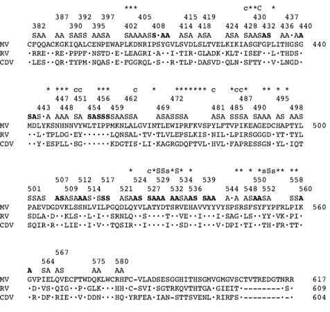
Measles virus (MV) enters cells either through the signaling lymphocyte activation molecule SLAM (CD150) expressed only in immune cells or through the ubiquitously expressed regulator of complement activation, CD46. To identify residues on the attachment protein hemagglutinin (H) essential for fusion support through either receptor, we devised a strategy based on analysis of morbillivirus H-protein sequences, iterative cycles of mutant protein production followed by receptor-based functional assays, and a novel MV H three-dimensional model. This model uses the Newcastle disease virus hemagglutinin-neuraminidase protein structure as a template. We identified seven amino acids important for SLAM- and nine for CD46 (Vero cell receptor)-induced fusion. The MV H three-dimensional model suggests (i) that SLAM- and CD46-relevant residues are located in contiguous areas in propeller beta-sheets 5 and 4, respectively; (ii) that two clusters of SLAM-relevant residues exist and that they are accessible for receptor contact; and (iii) that several CD46-relevant amino acids may be shielded from direct receptor contacts. It appears likely that certain residues support receptor-specific H-protein conformational changes. To verify the importance of the H residues identified with the cell-cell fusion assays for virus entry into cells, we transferred the relevant mutations into genomic MV cDNAs. Indeed, we were able to recover recombinant viruses, and we showed that these replicate selectively in cells expressing SLAM or CD46. Selectively receptor-blind viruses will be used to study MV pathogenesis and may have applications for the production of novel vaccines and therapeutics.


Figures

FIG. 1.
FIG. 1.
Sequences of parts of three morbillivirus H proteins and structure of the 60-block mutants produced. The MV, RV, and CDV sequences are shown. The MV H sequence is shown in full; dots are used in the RV and CDV sequences to indicate conserved residues. The three lines above the MV sequence are used to define the 60-block mutants. Two series of mutants were produced: the first included 16 blocks of conserved residues (in boldface), the second 44 blocks of divergent residues (in lightface). Alanine (A) was used to replace charged and polar residues, and serine (S) was used to replace apolar residues. The position of the first amino acid of each block mutant is indicated by an ordinal number. In the top line, the positions of the 56 single amino acid mutants are indicated with an asterisk or a letter. A capital “C” or “S” indicates residues whose mutation completely abolished CD46- or SLAM-dependent fusion support, respectively; a lowercase “c” or “s” indicates residues whose mutation caused strong or moderate reduction in specific receptor-dependent fusion support. All other mutants are indicated by asterisks.
FIG. 2.
FIG. 2.
Fusion efficiency of the H-protein mutants in Vero or CHO-SLAM cells. The 60 H-protein block mutants are represented as rectangles in the top half, and the 56 single residue mutants are shown as bars in the bottom half. A filled rectangle or bar indicates full fusion activity, a void rectangle or bar indicates no fusion activity, and two-thirds- and one-third-filled objects indicate intermediate fusion levels as defined in Materials and Methods. The mutants are drawn according to their ordinal position in the linear H sequence. Block mutants with strong receptor-mediated fusion differential are highlighted with asterisks (upper half); single-residue mutants with strong receptor-mediated fusion differential are indicated by the ordinal numbers corresponding to their positions (lower half).
FIG. 3.
FIG. 3.
Alignment of the MV H with the NDV HN protein sequences and secondary structure predictions used for modeling. The 18 secondary structure motifs identified with the program MASIA are indicated above the MV H sequence with a letter (a to r) and boxed. On the top line the results of the JPred structure prediction of H are shown. The secondary structure of the template is shown below the NDV sequence: arrowed lines indicate β-sheets and boxes indicate alpha-helical regions. The structures are color coded according to each of the six predicted “propeller” sheets from 1 to 6: yellow, red, cyan, pink, blue, and green, respectively.
FIG. 4.
FIG. 4.
New MV H-protein structural model showing both ribbon plot (A and B) and space-filling (C and D) representations. Panels A and C are a view of the protein from the top; in panels B and D the model was rotated 270° in a mathematically positive direction around the x axis. The same nomenclature as introduced for the NDV HN protein is used here. The globular head of the protein is predicted to consist of a superbarrel in which six β sheets (sheets 1 to 6) are arranged cyclically around an axis like the blades of a propeller and loops protrude from the top and lower surfaces of each “blade.” The color code used in panels A and B is the same as that used in Fig. 3. In panels C and D, all residues that have been mutated in blocks are shown in avocado green; all of the individually mutated amino acids are shown in light blue and have been numbered. Residues indicated in gray were not mutated.
FIG. 5.
FIG. 5.
Predicted location of all amino acids whose mutation reduces receptor-dependent fusion-support function on the new MV H-protein structural model. (A and C) Top views of the H molecule; (B and D) side views of the H molecule. (A and B) Residues whose mutation abolished SLAM-dependent fusion function are shown in red; those whose mutation strongly or moderately impaired SLAM-dependent fusion function are in gold. (C and D) The only residue whose mutation abolished CD46 (Vero cell)-dependent fusion is shown in pink; those whose mutation strongly or moderately impaired CD46-dependent fusion function are shown in dark blue. The ordinal number and chemical nature of all of the amino acids that selectively impaired fusion with any receptor are indicated. Residue V451, which strongly impaired CD46-dependent fusion, is located below the solvent-exposed surface.
FIG. 6.
FIG. 6.
Infection of Vero and B95a cells with selectively receptor-blind viruses. Green fluorescence emission of cells infected with different viruses (A) and analysis of H-protein production in the protein extracts of the same cells (B). Cells were infected with equivalent MOIs of the seven viruses indicated, photographed at 30 (Vero) or 48 (B95a) h postinfection, and lysed; protein extracts were prepared, and the expression of the H protein was analyzed by Western blot. All viruses had an identical genome, including the reporter protein GFP expressed from an additional transcription unit, and differed only in the H-protein amino acid(s) indicated. The H(wt)V451S,A527S protein, with two predicted additional glycosylation sites, migrates more slowly than all other proteins.

Similar articles

Cited by

References

    1. Altschul, S. F., T. L. Madden, A. A. Schaffer, J. Zhang, Z. Zhang, W. Miller, and D. J. Lipman. 1997. Gapped BLAST and PSI-BLAST: a new generation of protein database search programs. Nucleic Acids Res. 25:3389-3402. - PMC - PubMed
    1. Angel, J. B., P. Walpita, R. A. Lerch, M. S. Sidhu, M. Masurekar, R. A. DeLellis, J. T. Noble, D. R. Snydman, and S. A. Udem. 1998. Vaccine-associated measles pneumonitis in an adult with AIDS. Ann. Intern. Med. 129:104-106. - PubMed
    1. Bartz, R., U. Brinckmann, L. M. Dunster, B. Rima, V. Ter Meulen, and J. Schneider-Schaulies. 1996. Mapping amino acids of the measles virus hemagglutinin responsible for receptor (CD46) downregulation. Virology 224:334-337. - PubMed
    1. Borrow, P., and M. B. Oldstone. 1995. Measles virus-mononuclear cell interactions. Curr. Top. Microbiol. Immunol. 191:85-100. - PubMed
    1. Bucheit, A. D., S. Kumar, D. Grote, Y. Lin, V. von Messling, R. Cattaneo, and A. K. Fielding. 2003. An oncolytic measles virus engineered to enter cells through the CD20 antigen. Mol. Ther. 7:62-72. - PubMed

Publication types

MeSH terms

LinkOut - more resources