. 2004 Jan;78(1):320-8.
doi: 10.1128/jvi.78.1.320-328.2004.

The ORF2 protein of hepatitis E virus binds the 5' region of viral RNA

Affiliations

The ORF2 protein of hepatitis E virus binds the 5' region of viral RNA

Milan Surjit et al. J Virol. 2004 Jan.

Abstract

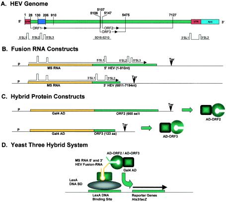
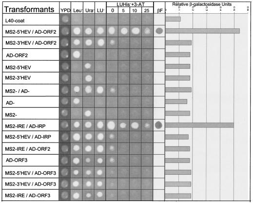
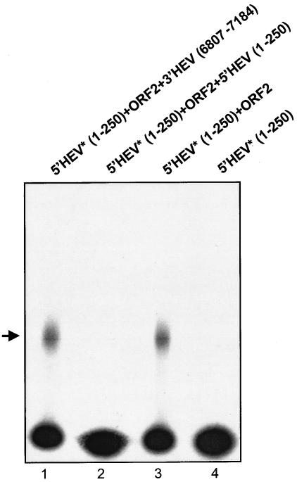
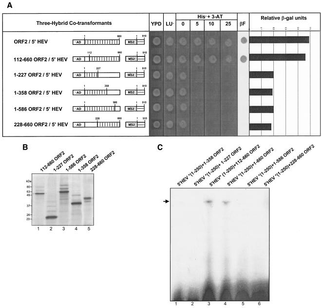
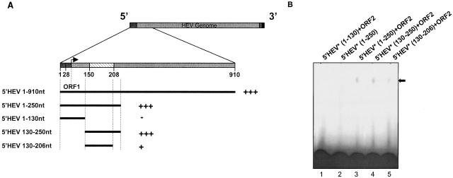
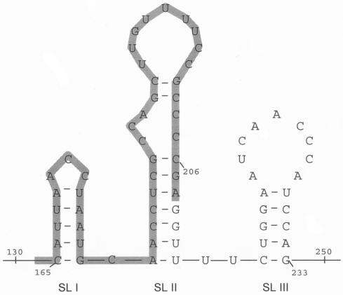
Hepatitis E virus (HEV) is a major human pathogen in much of the developing world. It is a plus-strand RNA virus with a 7.2-kb polyadenylated genome consisting of three open reading frames, ORF1, ORF2, and ORF3. Of these, ORF2 encodes the major capsid protein of the virus and ORF3 encodes a small protein of unknown function. Using the yeast three-hybrid system and traditional biochemical techniques, we have studied the RNA binding activities of ORF2 and ORF3, two proteins encoded in the 3' structural part of the genome. Since the genomic RNA from HEV has been postulated to contain secondary structures at the 5' and 3' ends, we used these two terminal regions, besides other regions within the genome, in this study. Experiments were designed to test for interactions between the genomic RNA fusion constructs with ORF2 and ORF3 hybrid proteins in a yeast cellular environment. We show here that the ORF2 protein contains RNA binding activity. The ORF2 protein specifically bound the 5' end of the HEV genome. Deletion analysis of this protein showed that its RNA binding activity was lost when deletions were made beyond the N-terminal 111 amino acids. Finer mapping of the interacting RNA revealed that a 76-nucleotide (nt) region at the 5' end of the HEV genome was responsible for binding the ORF2 protein. This 76-nt region included the 51-nt HEV sequence, conserved across alphaviruses. Our results support the requirement of this conserved sequence for interaction with ORF2 and also indicate an increase in the strength of the RNA-protein interaction when an additional 44 bases downstream of this 76-nt region were included. Secondary-structure predictions and the location of the ORF2 binding region within the HEV genome indicate that this interaction may play a role in viral encapsidation.


Figures

FIG. 1.
FIG. 1.
HEV genome and yeast three-hybrid constructs used to study RNA-protein interactions. (A) Genes and genome organization of HEV. Start sites for all three ORFs and both 3′ and 5′ UTRs are shown in red. A(n) represents the poly(A) tail (blue). Predicted stem-loop structures (SL) are shown schematically and numbered across the HEV genome. (B) Fusion RNA constructs designed to express fusion transcripts within the yeast cell. The MS2 RNA coding region (yellow) was cloned with two different HEV genomic regions (green). Predicted mRNA stem-loop structures are shown schematically as fusion transcripts. (C) Hybrid protein constructs to test the RNA binding activity of the ORF2 and ORF3 proteins of HEV using the yeast three-hybrid system. The schematic diagram shows the Gal4 Gal4 AD fused in frame to the ORF2 and ORF3 genes of HEV, thus expressing fusion proteins in yeast cells. (D) Schematic diagram of the yeast three-hybrid system showing the different fusion-RNA and hybrid-protein constructs being examined. P, promoter; Ter, terminator; SL, stem-loop structure.
FIG. 2.
FIG. 2.
Results from the three-hybrid analysis showing 5′ HEV genomic RNA interacting with the ORF2 protein. YPD, yeast extract peptone dextrose media (nonselective); Leu, Ura, LU represent SD-Leu (synthetic dextrose complete medium lacking leucine), SD-Ura (synthetic dextrose complete media lacking uracil), and SD-Leu Ura synthetic growth media. LUHis+3-AT (synthetic dextrose complete media lacking histidine, leucine, and uracil with 3-aminotrizole) represents SD-Leu Ura His synthetic medium with 0, 5, 10, and 25 mM 3-aminotrizole (3-AT) added. βF represents results from the β-galactosidase filter assay, and the bar graph represents relative β-galactosidase units from the liquid β-galactosidase assay. L40-coat is the untransformed yeast host strain. MS2-IRE/AD-IRP is the postive control used in the assay (26).
FIG. 3.
FIG. 3.
In vitro gel shift assay confirming the results of the yeast three-hybrid system. 5′ HEV RNA (nt 1 to 250) and 3′HEV RNA (nt 6807 to 7184) were 32P radiolabeled. ORF2 protein was produced using a coupled transcription-translation system. The arrow shows ORF2 protein bound to 32P-labeled RNA (lanes 2 and 3). Lanes 1 and 4 are negative controls. Asterisks refer to 32P labeled transcripts.
FIG. 4.
FIG. 4.
Competitor binding assay. The ORF2 protein used in this experiment is unlabeled. Lane 1 contains a nonspecific competitior, 3′ HEV RNA (nt 6807 to 7184). Lane 2 contains a 100-fold excess of unlabeled 5′HEV RNA (nt 1 to 250) transcript. Lanes 3 and 4 are positive and negative controls, respectively. Asterisks refer to 32P-labeled transcripts. Nonradioactive transcripts 3′ HEV RNA (nt 6807 to 7184) and 5′ HEV RNA (nt 1 to 250) were used at 100-fold higher molar concentrations.
FIG. 5.
FIG. 5.
Mapping the interaction domain for the ORF2 protein. (A) Amino acids 112 to 660 from the ORF2 protein are required for interaction with the 5′ HEV RNA (nt 1 to 910) region. Dotted boxes represent the AD regions which were fused in frame with the ORF2 protein (full length or deletions) shown in boxed regions with vertical lines. Checkered boxes show the MS2 regions fused with the 5′ HEV RNA (nt 1 to 910), shown as horizontal lines. Open boxes represent regions that were deleted from ORF2. The numbers above the boxed regions with vertical lines represent the first and last nucleotides of the regions included in the ORF2 deletion constructs. YPD, yeast extract peptone dextrose media (nonselective); LU, SD-Leu Ura synthetic growth medium; LUHis+3-AT, SD-Leu Ura His synthetic medium with 0, 5, 10, and 25 mM 3-AT added. βF represents results from the β-galactosidase filter assay, and the bar graph represents relative β-galactosidase units from the liquid β-galactose assay. (B) Control gel showing ORF2 deletions expressed using a coupled transcription-translation expression system. Major bands show the expressed protein of interest and correspond to their calculated molecular masses. Weaker bands in each lane show nonspecific translation of rabbit reticulocyte proteins. (C) EMSA showing ORF2(112-660) interacting with the 5′ genomic region of HEV. Asterisks refer to 32P-labeled transcript.
FIG. 6.
FIG. 6.
Mapping the interaction domain for the 5′ HEV RNA. (A) Mapping of the interaction domain of the 5′ HEV RNA (nt 1 to 910) region. The hatched box represents the alphavirus consensus sequence. Plus signs shows a summarized result of the yeast three-hybrid interactions. (B) EMSA for the different RNA deletions from the 5′ HEV genome. Asterisks refer to 32P-labeled transcript.
FIG. 7.
FIG. 7.
Secondary-structure prediction of the 5′ HEV genomic region from nt 130 to 250. Numbers correspond to the numbers on the HEV genome. SL I, SL II, and SL III represent the three stem-loop structures shown in the figure. The highlighted region represents the 51-nt conserved region from alphaviruses.

References

    1. Aye, T. T., T. Uchida, X. Z. Ma, F. Iida, T. Shikata, H. Zhuang, and K. M. Win. 1992. Complete nucleotide sequence of a hepatitis E virus isolated from the Xinjiang epidemic (1986-1988) of China. Nucleic Acids Res. 20:3512. - PMC - PubMed
    1. Berke, T., and D. O. Matson. 2000. Reclassification of the Calciviridae into distinct genera and exclusion of hepatitis E virus from the family on the basis of comparative phylogenetic analysis. Arch. Virol. 145:1421-1436. - PubMed
    1. Bradley, D. W. 1990. Enterically-transmitted non-A, non-B hepatitis. Br. Med. Bull. 46:442-461. - PubMed
    1. Bradley, D. W., and M. A. Purdy. 1994. Molecular and serological characteristics of hepatitis E virus, p 42-45. In K. Nishioka, H. Suzuki, S. Mishiro, et al. (ed.) Viral hepatitis and liver disease. Springer-Verlag, Tokyo, Japan.
    1. Guarente, L. 1983. Yeast promoters and lacZ fusions designed to study expression of cloned genes in yeast. Methods Enzymol. 101:181-191. - PubMed

Publication types