. 2004 Jan;78(1):329-39.
doi: 10.1128/jvi.78.1.329-339.2004.

Characterization of the interaction between P143 and LEF-3 from two different baculovirus species: Choristoneura fumiferana nucleopolyhedrovirus LEF-3 can complement Autographa californica nucleopolyhedrovirus LEF-3 in supporting DNA replication

Affiliations

Characterization of the interaction between P143 and LEF-3 from two different baculovirus species: Choristoneura fumiferana nucleopolyhedrovirus LEF-3 can complement Autographa californica nucleopolyhedrovirus LEF-3 in supporting DNA replication

Tricia Chen et al. J Virol. 2004 Jan.

Abstract

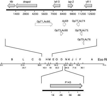
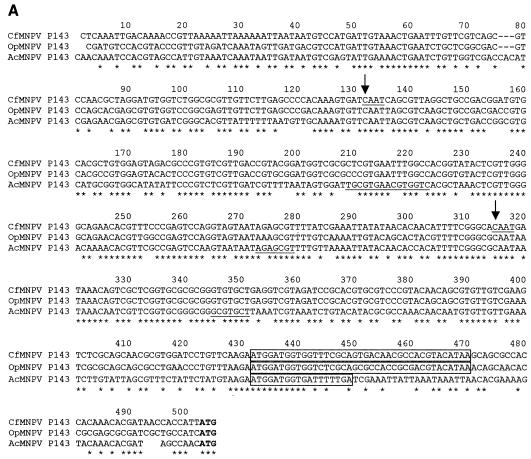
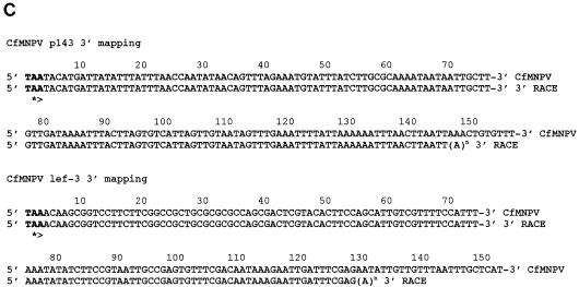
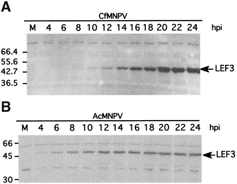
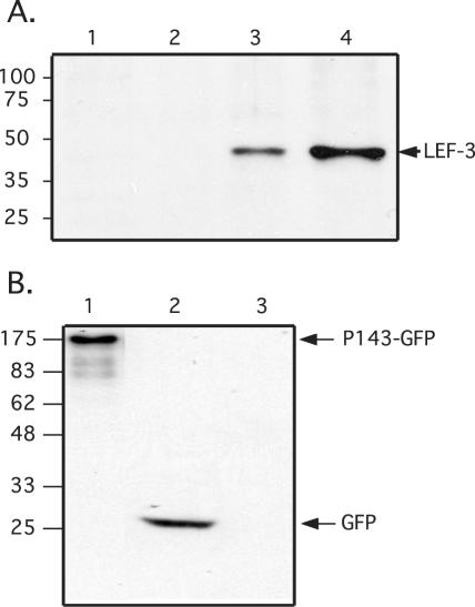
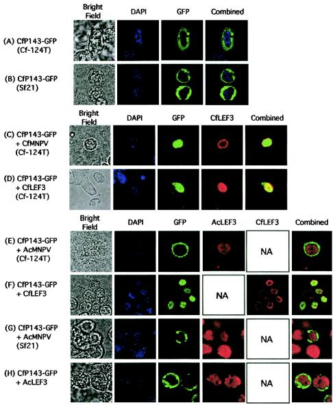
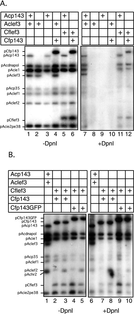
The baculovirus protein P143 is essential for viral DNA replication in vivo, likely as a DNA helicase. We have demonstrated that another viral protein, LEF-3, first described as a single-stranded DNA binding protein, is required for transporting P143 into the nuclei of insect cells. Both of these proteins, along with several other early viral proteins, are also essential for DNA replication in transient assays. We now describe the identification, nucleotide sequences, and transcription patterns of the Choristoneura fumiferana nucleopolyhedrovirus (CfMNPV) homologues of p143 and lef-3 and demonstrate that CfMNPV LEF-3 is also responsible for P143 localization to the nucleus. We predicted that the interaction between P143 and LEF-3 might be critical for cross-species complementation of DNA replication. Support for this hypothesis was generated by substitution of heterologous P143 and LEF-3 between two different baculovirus species, Autographa californica nucleopolyhedrovirus and CfMNPV, in transient DNA replication assays. The results suggest that the P143-LEF-3 complex is an important baculovirus replication factor.


Figures

FIG. 1.
FIG. 1.
Location of identifiable ORFs in the sequenced region of p143 and lef-3. The regions of CfMNPV that were sequenced to identify the lef-3 (above) and p143 (below) genes are indicated and oriented on the CfMNPV genome EcoRI restriction fragment map. The presence of ORFs with predicted functions is indicated as filled arrows above the scale in base pairs. ORFs with homologues in OpMNPV and AcMNPV and their numbers but no specific function are indicated as open arrows below the scale line.
FIG. 2.
FIG. 2.
Expression and mapping of P143 and LEF-3 transcripts. The upper diagrams (A) show the orientation of the mRNAs, the open reading frames, and the location of the strand-specific riboprobes used in the Northern analysis for the CfMNPV p143 and lef-3 genes. Also shown are the names and locations of the primers used in the 5′ and 3′ RACE analysis to map the 5′ and 3′ ends of the p143 and lef-3 mRNAs. (B) Poly(A)+ RNA, prepared from CfMNPV-infected Cf124T cells at the times indicated was resolved by 0.6% agarose gel electrophoresis. Blots of these gels were probed with strand-specific riboprobes corresponding to the p143 and lef-3 genes. Similarly prepared poly(A)+ RNA from mock-infected cells was included as controls (M). The exposures were long, to enable the detection of virus-specific mRNA at the early time point (6 h postinfection). The sizes of the detectable transcripts are indicated on the right side of each blot. (C) PCR products generated from the 5′and 3′ RACE analysis of the p143 and lef-3 mRNA were separated on agarose gels. Sequence analysis of these products revealed the 5′ transcription start site and 3′ polyadenylation site for each gene (shown in Fig. 3).
FIG. 3.
FIG. 3.
Promoter sequence for p143 and lef-3. An alignment of the promoter regions of the p143 (A) and lef-3 (B) genes from CfMNPV, OpMNPV, and AcMNPV is shown. TATA-box-like sequences are shaded, the location of published transcription start sites are underlined, minicistron coding regions are boxed and the translation start codons are in bold. The locations of the transcription start sites for the CfMNPV p143 and lef-3 genes, as determined by sequence analysis of PCR products, are shown with arrows. (C) The sequences of the 3′ ends of the p143 and lef-3 mRNAs as determined by 3′ RACE and sequence analysis of PCR products are shown below the appropriate genomic sequence.
FIG. 3.
FIG. 3.
Promoter sequence for p143 and lef-3. An alignment of the promoter regions of the p143 (A) and lef-3 (B) genes from CfMNPV, OpMNPV, and AcMNPV is shown. TATA-box-like sequences are shaded, the location of published transcription start sites are underlined, minicistron coding regions are boxed and the translation start codons are in bold. The locations of the transcription start sites for the CfMNPV p143 and lef-3 genes, as determined by sequence analysis of PCR products, are shown with arrows. (C) The sequences of the 3′ ends of the p143 and lef-3 mRNAs as determined by 3′ RACE and sequence analysis of PCR products are shown below the appropriate genomic sequence.
FIG. 4.
FIG. 4.
Temporal expression of CfMNPV LEF-3 in infected cells. Cf124T cells, infected with CfMNPV, were harvested at the indicated times after infection (A). Whole-cell extracts were resolved by SDS-10% PAGE, blotted onto nitrocellulose filters, and then probed with polyclonal antibodies against CfMNPV LEF-3. CfMNPV LEF-3 was first clearly detectable at 8 h postinfection. For comparison, a similar blot of extracts prepared from AcMNPV-infected Sf21 cells and probed with LEF-3-specific polyclonal antibody is shown. (B) AcMNPV LEF-3 was first detectable at 4 h postinfection.
FIG. 5.
FIG. 5.
Expression of LEF-3 and P143-GFP following transfection or infection and detected by immunoblotting. (A) Whole-cell extracts (5 × 10⁴ cells per lane) were prepared from mock-infected Sf21 cells (lane 1), mock-infected Cf124T cells (lane 2), CfMNPV-infected Cf124T cells (lane 3), or pIE1hrCflef-3-transfected Sf21 cells (lane 4) at 24 h posttransfection or postinfection. The extracts were analyzed by SDS-10% PAGE, transferred to a nitrocellulose membrane and probed with LEF-3-specific polyclonal antibody. The relative mobility of molecular weight markers is shown on the left and the immunoreactive proteins are labeled on the right. (B) Whole-cell extracts (5 × 10⁴ cells per lane) were prepared from pIE1hrCfp143GFP-transfected Sf21 cells (lane 1), pAcGFP-transfected Sf21 cells (lane 2), and mock-transfected Sf21 cells (lane 3) harvested at 24 h posttransfection. Whole-cell extracts were analyzed by SDS-11.25% PAGE, transferred to a nitrocellulose membrane and probed with anti-GFP monoclonal antibody. The relative mobility of the molecular weight markers is shown on the left and the immunoreactive proteins are labeled on the right.
FIG. 6.
FIG. 6.
Intracellular localization of P143 and LEF-3 following transfection detected by immunofluorescence. Cf124T (A, C, D, and E) or Sf21 (B and F to H) cells, transfected with plasmids expressing CfMNPV P143 fused to GFP (pIE1hrCfp143GFP) (A to H), CfMNPV LEF-3 (pIE1hrCflef-3) (D and H), or AcMNPV LEF-3 (pAcLEF-3) (G), were mock infected or infected with CfMNPV (C) or AcMNPV (E and F). At 24 h posttransfection, the cells were either observed directly for GFP fluorescence or were also processed for immunofluorescence using antibodies directed against CfMNPV LEF-3 (CfLEF3) or AcMNPV LEF-3 (AcLEF3). Nuclear DNA was stained with DAPI. Only infection with CfMNPV or cotransfection with CfMNPV LEF-3 resulted in nuclear GFP fluorescence from CfMNPV P143-GFP.
FIG. 7.
FIG. 7.
Transient plasmid DNA replication in the presence of heterologous P143 and LEF-3 proteins. Sf21 cells were transfected with a collection of plasmids, which together expressed the AcMNPV genes necessary for plasmid DNA replication (ie-1, dnapol, lef-1, lef-2, p35, pe38, and ie-2) except p143 and lef-3. In separate transfections, this library was supplemented with plasmids expressing the AcMNPV p143 (Acp143), AcMNPV lef-3 (Aclef3), CfMNPV p143 (Cfp143) or CfMNPV lef-3 (Cflef3) genes. Following incubation for 48 h, total intracellular DNA was prepared and digested with EcoRI (−DpnI) to linearize the plasmids or with EcoRI and DpnI (+DpnI) to detect replicated plasmid DNA. Southern blots of these restriction digestion DNA preparations were probed with labeled pUC19 DNA. Replication of input plasmid DNA was detected in the presence of plasmidsexpressing AcMNPV P143 and LEF-3, CfMNPV P143 and LEF-3, and AcMNPV P143 and CfMNPV LEF-3 (A). Similar assays were also done with a plasmid expressing the CfMNPV P143-GFP fusion protein (B). This protein also supported plasmid DNA replication in the presence of CfMNPV LEF-3.

Similar articles

Cited by

References

    1. Ahrens, C. H., C. Carlson, and G. F. Rohrmann. 1995. Identification, sequence, and transcriptional analysis of lef-3, a gene essential for Orgyia pseudotsugata baculovirus DNA replication. Virology 210:372-382. - PubMed
    1. Ahrens, C. H., and G. F. Rohrmann. 1996. The DNA polymerase and helicase genes of a baculovirus of Orgyia pseudotsugata. J. Gen. Virol. 77:825-837. - PubMed
    1. Argaud, O., L. Croizier, M. López-Ferber, and G. Croizier. 1998. Two key mutations in the host-range specificity domain of the p143 gene of Autographa californica nucleopolyhedrovirus are required to kill Bombyx mori larvae. J. Gen. Virol. 79:931-935. - PubMed
    1. Bell, S. P., and A. Dutta. 2002. DNA replication in eukaryotic cells. Annu. Rev. Biochem. 71:333-374. - PubMed
    1. Bideshi, D. K., and B. A. Federici. 2000. The Trichoplusia ni granulovirus helicase is unable to support replication of Autographa californica multicapsid nucleopolyhedrovirus in cells and larvae of T. ni. J. Gen. Virol. 81:1593-1599. - PubMed

Publication types

MeSH terms

Associated data

LinkOut - more resources