. 2004 Jan;78(1):482-90.
doi: 10.1128/jvi.78.1.482-490.2004.

Cultured peripheral neuroglial cells are highly permissive to sheep prion infection

Affiliations

Cultured peripheral neuroglial cells are highly permissive to sheep prion infection

Fabienne Archer et al. J Virol. 2004 Jan.

Abstract

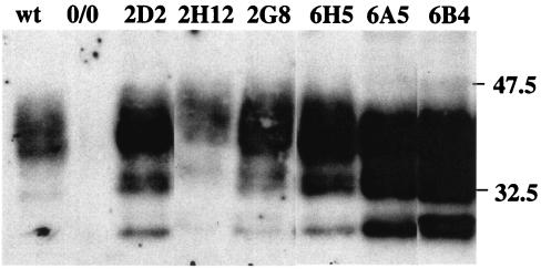
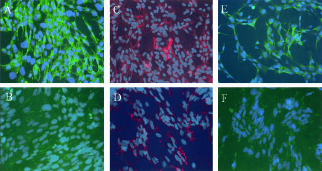
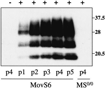
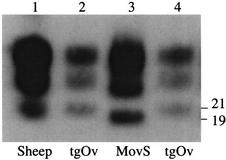
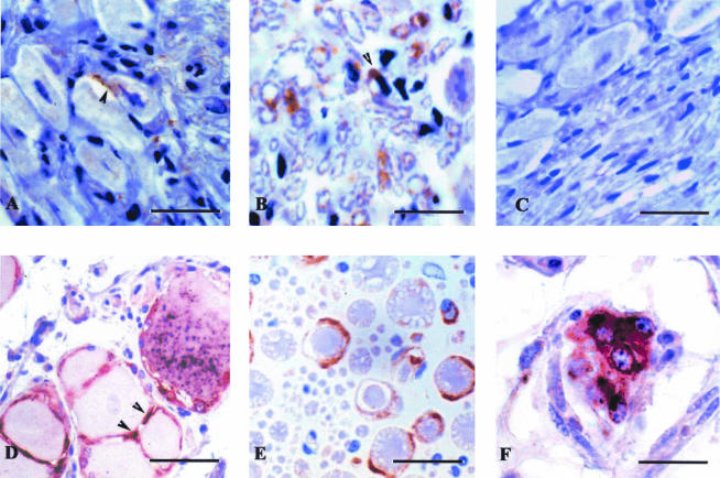
Transmissible spongiform encephalopathies arise as a consequence of infection of the central nervous system (CNS) by prions. Spreading of the infectious agent through the peripheral nervous system (PNS) may represent a crucial step toward CNS neuroinvasion, but the modalities of this process have yet to be clarified. Here we provide further evidence that PNS glial cells are likely targets for infection by prions. Glial cell clones originating from dorsal root ganglia of transgenic mice expressing ovine PrP (tgOv) and simian virus 40 T antigen were found to be readily infectible by sheep scrapie agent. This led us to establish two stable cell lines that exhibited features of Schwann cells. These cells were shown to sustain an efficient and stable replication of sheep prion based on the high level of accumulation of abnormal PrP and infectivity in exposed cultures. We also provide evidence for abnormal PrP deposition in peripheral neuroglial cells from scrapie-infected tgOv mice and sheep. These findings have potential implications in terms of designing new cell systems permissive to prions and of peripheral pathobiology of prion infections.


Figures

FIG. 1.
FIG. 1.
PrPc expression in neuroglial cell clones. Six clones derived from DRG of ovine PrP transgenic mice (Table 1) were analyzed for PrPc with 4F2 antibody. A Western blot performed on lysates from cultures at 11 to 15 passages postexplantation of the indicated clones and from primary glial cell cultures of C57BL6 (wild type [wt]) or PrP0/0 (0/0) mice is shown (10 μg of protein/lane, equivalent to 3 × 10⁴ cells). Molecular masses (in kilodaltons) are indicated to the right of the blot.
FIG. 2.
FIG. 2.
Expression of Schwann cell markers in MovS cells. Immunohistochemical characterization of MovS6 (A, C, and E) and MovS2 cells (B, D, and F). p75-NGFR (A and B) is expressed by MovS6 cells but not MovS2 cells. GalC (C and D) is expressed by a minority of cells in both clones. GFAP (E and F) is not expressed by MovS2 but is expressed by a subpopulation of MovS6 cells.
FIG. 3.
FIG. 3.
Dynamics of PrPres accumulation in MovS6 cells following exposure to sheep prion. PK-treated cell lysates (500 μg of protein) from cultures at the indicated passage p.i. (p1 to p5) were analyzed by Western blotting with 2D6 antibody. MovS6 and MS0/0 cultures (12th subpassage) were incubated for 4 days with 2.5% brain homogenate prepared from scrapie-diseased (+) or control (−) tgOv mice. MOI, ∼20 ID50 U/cell.
FIG. 4.
FIG. 4.
In situ detection of PrPsc in MovS2 cells infected with sheep prion. (Top) Slices of paraffin-embedded, mock-infected (left) and infected cells (13 passages p.i.) (right) were PK treated and immunolabeled with 8G8 antibody and peroxidase (see Materials and Methods). PrPres deposits are visible at the intracellular and plasma membrane (inset) levels. (Bottom) Fixed cell monolayers were treated with guanidine thiocyanate and immunolabeled with ICSM18 antibody. In infected cultures (panels 2 and 3), most cells show a punctuate fluorescence not seen in mock-infected cultures (panel 1) and are thus assumed to reflect abnormal PrP accumulation. Magnifications, ×200 and ×400.
FIG. 5.
FIG. 5.
Reversible change of the molecular profile of sheep scrapie agent propagated in MovS cells. PK-digested material (50 μg) derived from brain tissues (500 μg) or a cell culture (105 cells) infected by the same source of scrapie agent was analyzed by Western blotting (2D6 antibody). The original PG127 scrapie isolate (lane 1) was passaged once in tgOv mice (lane 2) and then propagated for 9 subpassages in MovS6 cells (lane 3) and reinoculated into tgOv mice (lane 4). Molecular masses (in kilodaltons) are indicated to the right of the figure.
FIG. 6.
FIG. 6.
Infection of MovS cells by exposure to low infectious dose in culture or by in vivo infection within the brain of a scrapie-diseased mouse. (A) PrPres detection (ICSM18 antibody) in MovS6 cultures serially passaged (p1 to p5) following a 6-day incubation with 0.5 ml of 2.5% infectious tgOv brain homogenate at the indicated dilutions. (B) Cell blot assay on MovS2 cells recovered from the brain of infected tgOv mice drafted with uninfected cells. Two tgOv mice at the onset of the clinical phase were inoculated intracerebrally with 3 × 10⁶ cells, and 4 days later their brains were removed and explanted into culture. Cell colonies formed within 5-day-old primary cultures were scraped, individually seeded on coverslips, and analyzed for PrPres 1 week later (8G8 antibody). Cell colonies derived from infected brain inoculated with Mov cells just before explantation into culture (−) or from a persistently infected culture (+) were used as negative and positive controls, respectively.
FIG. 7.
FIG. 7.
Detection of PrPres in PNS glial cells derived from infected tgOv mice. Primary cultures were established from DRG of terminally diseased animals inoculated by the peritoneal route or of control animals (−). PK-resistant PrP was revealed in cell lysates (Western blotting) (left) or monolayers (cell blotting) (right) from cultures at the indicated passage level (p0, 10 days postexplantation). A cell blot on a monolayer from a persistently infected Mov culture (+) is shown for comparison. Molecular mass (in kilodaltons) is indicated on the left.
FIG. 8.
FIG. 8.
PrPsc accumulation in DRG from scrapie-diseased mice and sheep. DRG from intraperitoneally inoculated tgOv mice (A and B) and naturally scrapie-diseased sheep (D to F) were formalin fixed, paraffin embedded, and immunolabeled for PrPsc (8G8 antibody). PrPsc deposits involve both neurons and glial cells: (i) satellite cells in panels A (diaminobenzidine [DAB] revelation, brown end product; bar, 15 μm) and D (5-bromo-4-chloro-3-indolylphosphate [BCIP]-nitroblue tetrazolium [NBT] revelation, black end product; bar, 25 μm) and (ii) axon ensheathing Schwann cells in panels B and E (DAB revelation; bar, 25 μm). Double labeling for PrPsc (NBT-BCIP revelation, black end product) and GFAP (3-amino-9-ethylcarbazole revelation, red end product) applied to sheep DRG shows PrPsc in GFAP-positive satellite cells (panel D) and interstitial, isolated Schwann cells (panel F; bar, 15 μm). No PrP labeling is seen in DRG from mock-infected mice (panel C; bar, 15 μm).

References

    1. Aguzzi, A. 1997. Neuro-immune connection in spread of prions in the body? Lancet 349:742-743. - PubMed
    1. Andreoletti, O., P. Berthon, E. Levavasseur, D. Marc, F. Lantier, E. Monks, J. M. Elsen, and F. Schelcher. 2002. Phenotyping of protein-prion (PrPsc)-accumulating cells in lymphoid and neural tissues of naturally scrapie-affected sheep by double-labeling immunohistochemistry. J. Histochem. Cytochem. 50:1357-1370. - PubMed
    1. Antoine, J. C., J. L. Laplanche, J. F. Mosnier, P. Beaudry, J. Chatelain, and D. Michel. 1996. Demyelinating peripheral neuropathy with Creutzfeldt-Jakob disease and mutation at codon 200 of the prion protein gene. Neurology 46:1123-1127. - PubMed
    1. Bartz, J. C., A. E. Kincaid, and R. A. Bessen. 2003. Rapid prion neuroinvasion following tongue infection. J. Virol. 77:583-591. - PMC - PubMed
    1. Bartz, J. C., A. E. Kincaid, and R. A. Bessen. 2002. Retrograde transport of transmissible mink encephalopathy within descending motor tracts. J. Virol. 76:5759-5768. - PMC - PubMed

Publication types