

doi: 10.1128/jvi.78.1.513-519.2004.
Chromosome-protein interactions in polyomavirus virions
Affiliations
- PMID: 14671132
- PMCID: PMC303386
- DOI: 10.1128/jvi.78.1.513-519.2004
Item in Clipboard
Chromosome-protein interactions in polyomavirus virions
J Virol.
2004 Jan.
Abstract
In this work, we sought to determine whether the components of the murine polyomavirus capsid establish specific interactions with the minichromosome encapsidated into the mature viral particles by using the cis-diamminedichloroplatinum(II) cross-linking reagent. Our data indicated that VP1, but not minor capsid proteins, interacts with the viral genome in vivo. In addition, semiquantitative PCR assays performed on cross-linked DNA complexes revealed that VP1 binds to all regions of the viral genome but significantly more to the regulatory region. The implications of such an interaction for viral infectivity are discussed.
Figures
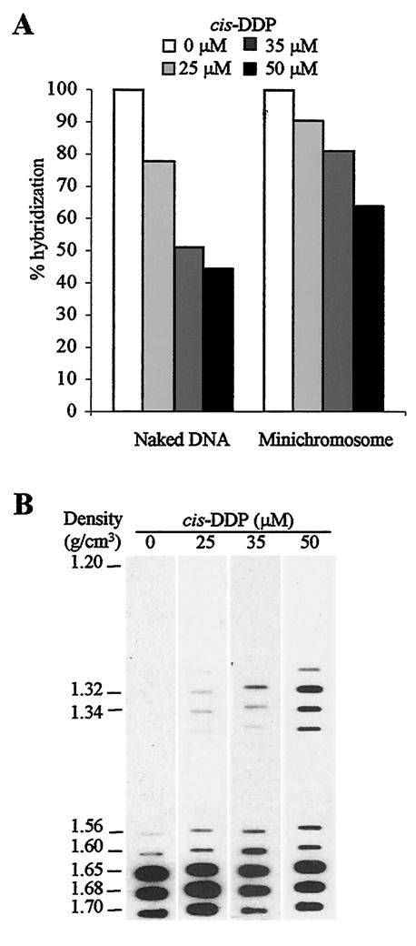
cis-DDP cross-links encapsidated minichromosomes. (A) cis-DDP penetrates viral particles. Naked viral DNA and viral particles were incubated with or without cis-DDP at different concentrations, and treated DNA was analyzed by Southern blotting with the whole Py genome used as a probe. The level of hybridization was quantified by phosphorimager analysis. One hundred percent hybridization corresponds to the level of hybridization detected in cis-DDP-untreated samples. (B) Effect of cis-DDP concentration on the recovery of DNA-protein complexes. Viral particles were incubated with different concentrations of cis-DDP. After virus dissociation, samples were ultracentrifuged in denaturing CsCl gradients. The distribution of viral DNA in the fractions of the gradient was analyzed by slot blotting with the whole Py genome used as a probe.
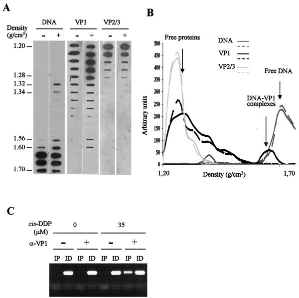
VP1 binds viral genome in mature particles. Qualitative (A) and quantitative (B) densitometry analysis of the distribution of viral DNA and capsid proteins VP1 and VP2/3 in the fractions of the gradient. Virions treated with (+) or without (−) the cis-DDP reagent are indicated at the top of each lane in panel A. Aliquots of each fraction were analyzed by slot blotting. Viral DNA was detected by hybridization as described in the legend to Fig. 1. Viral proteins were detected by using monoclonal antibodies directed against VP1 or VP2/3 (13) followed by incubation with horseradish peroxidase-conjugated secondary antibodies and an enhanced chemiluminescence reaction. Solid and broken lines represent cis-DDP-treated and cis-DDP-untreated virions, respectively, in panel B. (C) VP1 is associated with the viral genome. Complexes containing fractions of cis-DDP-treated (35 μM) or cis-DDP-untreated (0 μM) virions were immunoprecipitated with (+) or without (−) anti-VP1 antibodies. After reversal of cross-links, the DNA purified from the immunoprecipitated (IP) and immunodepleted (ID) samples was used as a template in PCRs (with primer pair 9) to amplify the Py origin of replication region.
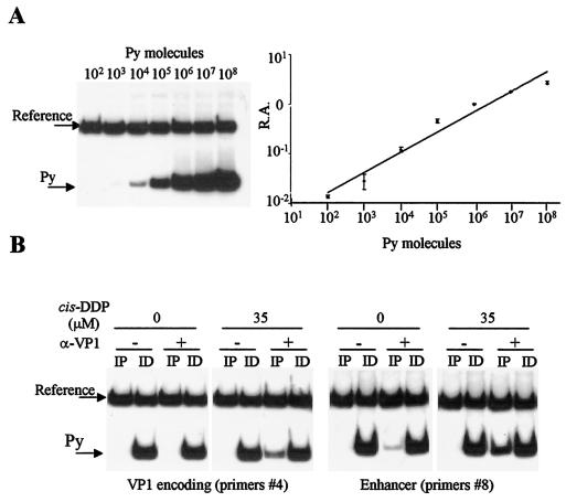
Experimental design of semiquantitative PCR analysis of VP1 cross-linked DNA fragments. (A) The relative amplification value (R.A.), measured as the ratio of amplification of Py to amplification of reference sequences, is a function of the initial amount of Py molecules present in the samples. (B) Representative PCR experiments performed in the presence of [α-P32]ATP with primer pairs 4 and 8 amplifying the VP1-encoding and enhancer regions, respectively.
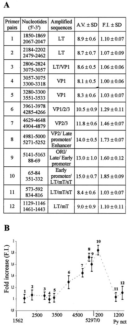
Semiquantitative PCR analysis of VP1 cross-linked DNA fragments. (A) Description of the primer pairs used to amplify target Py sequences. For a statistical analysis, each PCR was done in triplicateand repeated at least six times. The relative amplification value (A.V.) was calculated for each primer pair as the ratio of (Py/ref)IP to [(Py/ref)IP + (Py/ref)ID]. The fold increase (F.I.) value was calculated as the ratio of the amplified value of each primer pair to the amplified value of primer pair 4 (lowest amplified value). ORI, origin of replication; LT, large T antigen; mT, middle T antigen; sT, small T antigen. (B) Graphical representation of the semiquantitative PCR analysis. The numbering of the Py genome starts at the EcoRI site (nt 1562). nct, nucleotide.
Similar articles
-
VP1, the major capsid protein of the mouse polyomavirus, binds microtubules, promotes their acetylation and blocks the host cell cycle.FEBS J. 2017 Jan;284(2):301-323. doi: 10.1111/febs.13977. Epub 2017 Jan 9. FEBS J. 2017. PMID: 27885808
-
Nuclear localization of avian polyomavirus structural protein VP1 is a prerequisite for the formation of virus-like particles.J Virol. 2004 Jan;78(2):930-7. doi: 10.1128/jvi.78.2.930-937.2004. J Virol. 2004. PMID: 14694124 Free PMC article.
-
Possible role for cellular karyopherins in regulating polyomavirus and papillomavirus capsid assembly.J Virol. 2008 Oct;82(20):9848-57. doi: 10.1128/JVI.01221-08. Epub 2008 Aug 13. J Virol. 2008. PMID: 18701594 Free PMC article.
-
Immunotherapeutic polyoma and human papilloma virus-like particles.Immunotherapy. 2009 Mar;1(2):303-12. doi: 10.2217/1750743X.1.2.303. Immunotherapy. 2009. PMID: 20635947 Review.
-
Microtubules in Polyomavirus Infection.Viruses. 2020 Jan 18;12(1):121. doi: 10.3390/v12010121. Viruses. 2020. PMID: 31963741 Free PMC article. Review.
Cited by
-
Immune sensing of mouse polyomavirus DNA by p204 and cGAS DNA sensors.FEBS J. 2021 Oct;288(20):5964-5985. doi: 10.1111/febs.15962. Epub 2021 May 26. FEBS J. 2021. PMID: 33969628 Free PMC article.
-
A polyomavirus peptide binds to the capsid VP1 pore and has potent antiviral activity against BK and JC polyomaviruses.Elife. 2020 Jan 21;9:e50722. doi: 10.7554/eLife.50722. Elife. 2020. PMID: 31960795 Free PMC article.
-
How viruses use the endoplasmic reticulum for entry, replication, and assembly.Cold Spring Harb Perspect Biol. 2013 Jan 1;5(1):a013250. doi: 10.1101/cshperspect.a013250. Cold Spring Harb Perspect Biol. 2013. PMID: 23284050 Free PMC article. Review.
References
-
- Blasquez, V., A. Stein, C. Ambrose, and M. Bina. 1986. Simian virus 40 protein VP1 is involved in spacing nucleosomes in minichromosomes. J. Mol. Biol. 191:97-106. - PubMed
-
- Brabec, V., and Z. Balcarova. 1993. Restriction-enzyme cleavage of DNA modified by platinum(II) complexes. Eur. J. Biochem. 216:183-187. - PubMed
-
- Buckler-White, A. J., G. W. Humphrey, and V. Pigiet. 1980. Association of polyoma T antigen and DNA with the nuclear matrix from lytically infected 3T6 cells. Cell 22:37-46. - PubMed
Publication types
MeSH terms
Substances
LinkOut - more resources
Full Text Sources
