. 2003 Dec 23;100(26):15619-24.
doi: 10.1073/pnas.2635658100. Epub 2003 Dec 11.

p57(Kip2) cooperates with Nurr1 in developing dopamine cells

Affiliations

p57(Kip2) cooperates with Nurr1 in developing dopamine cells

Bertrand Joseph et al. Proc Natl Acad Sci U S A. .

Abstract

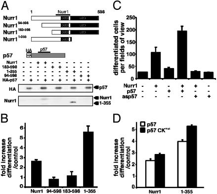
Cyclin-dependent kinase inhibitors of the Cip/Kip family play critical roles in regulating cell proliferation during embryogenesis. However, these proteins also influence cell differentiation by mechanisms that have remained unknown. Here we show that p57Kip2 is expressed in postmitotic differentiating midbrain dopamine cells. Induction of p57Kip2 expression depends on Nurr1, an orphan nuclear receptor that is essential for dopamine neuron development. Moreover, analyses of p57Kip2 gene-targeted mice revealed that p57Kip2 is required for the maturation of midbrain dopamine neuronal cells. Additional experiments in a dopaminergic cell line demonstrated that p57Kip2 can promote maturation by a mechanism that does not require p57Kip2-mediated inhibition of cyclin-dependent kinases. Instead, evidence indicates that p57Kip2 functions by a direct protein-protein interaction with Nurr1. Thus, in addition to its established function in control of proliferation, these results reveal a mechanism whereby p57Kip2 influences postmitotic differentiation of dopamine neurons.


Figures

Fig. 1.
Fig. 1.
p57Kip2 expression is detected in differentiating DA cells in vitro and in vivo. (A) Dox-induced Nurr1 induces p21Cip1 and p57Kip2 mRNA expression. Nurr1, p21Cip1, p27Kip1, and p57Kip2 mRNA content was determined by RT-PCR in MN9D Nurr1Tet-On cells. cDNA-encoding p21Cip1, p27Kip1, and p57Kip2 were used as PCR controls. Sagittal sections of E13.5 wild-type mouse embryos showing Nurr1 (B), p57Kip2 (C), and TH (D) mRNA expression by in situ hybridization analysis. Images on the right are close-ups of the ventral midbrain. Nurr1 was expressed in a broad domain in the ventral midbrain as expected, whereas p57Kip2 mRNA was detected in a more restricted domain coinciding with TH mRNA expression. (EG) Nurr1 (E) and p57Kip2 (F) immunoreactivity in confocal images of coronal midbrain sections. Overlay is shown in G. (Scale bars: BD Insets, 100 μm; EG, 50 μm.)
Fig. 2.
Fig. 2.
p57Kip2 expression is down-regulated in the Nurr1 mutant midbrain. Coronal sections of wild-type (A, C, E, and G) and Nurr1-null mutant (B, D, F, and H) ventral midbrain at E13.5 showing in situ hybridization detecting p57Kip2 (A and B), Nurr1 (C and D), TH (E and F), and En1 (G and H) mRNAs. In the Nurr1 mutant midbrain, expression of p57Kip2 is exclusively down-regulated in the mantle zone (mz) but not in the Nurr1-negative ventricular zone (vz) (B). In the wild type, Nurr1, TH, and En1 are expressed in the mantle zone (C, E, and G). Nurr1 and TH were not detected in the Nurr1 mutant ventral midbrain (D and F), whereas expression of En1 remains expressed in Nurr1-deficient cells (H). (Scale bar, 100 μm.)
Fig. 3.
Fig. 3.
p57Kip2 enhances DA cell differentiation in vitro and interacts directly with Nurr1. (A) MN9D cells were cotransfected with enhanced GFP (EGFP) expression vector together with expression vectors for Nurrl, p57Kip2, or both. The number of differentiated EGFP-expressing cells was counted 3 days after transfection as described (22). Average values of quadruplicates are shown. Error bars represent standard deviation. (B) p57Kip2 interacts with Nurr1. HEK 293 cells were transfected with the empty expression vector, expression vectors encoding Flag-Nurr1, HA-p57Kip2, or both. Nuclear cell extracts from transfected cells were subjected to immunoblotting with anti-HA or anti-Flag antibodies (Top). Nuclear cell extracts were immunoprecipitated by using anti-Nurr1 (Middle) or anti-p57Kip2 (Bottom) antibodies, and immune complexes were subjected to immunoblotting by using anti-HA or anti-Flag antibodies. (C) p57Kip2 interacts with Nurr1 in extracts from the embryonal ventral midbrain. Total cell extracts were prepared from rat E15 ventral midbrain and immunoprecipitated by using anti-IgG control (left lane) or anti-Nurr1 antibodies (center lane). Immune complexes were subjected to immunoblotting by using anti-p57Kip2 antibodies. Nuclear cell extract from HEK 293 cells transfected with expression vectors encoding p57Kip2 was used as control (right lane). (D) p57Kip2 and Nurr1 proteins interact in transfected cells. The interaction between p57Kip2 and Nurr1 was confirmed by using a mammalian two-hybrid assay. HEK 293 cells were transfected with expression vectors VP16-p57Kip2 and Gal4DBD-Nurr1 (1–262). These vectors were transfected either alone or as indicated in the figure together with a luciferase reporter gene driven by four UAS Gal4-binding sites. Relative light units (RLU) were computed after normalization to β-galactosidase activities. Gal4DBD-Nurr1 (1–262) activates the reporter gene due to the presence of a transactivation domain within the Nurr1 amino-terminal domain. Activation is strongly enhanced by cotransfection of VP16-p57Kip2. (E) A gel-mobility shift assay demonstrated that in vitro transcribed and translated HA-p57Kip2 formed complexes with in vitro translated Nurr1 bound to a 32P-labeled NBRE DNA probe. The positions of Nurr1 (shift) and Nurr1/p57Kip2 complexes (super shift) bound to NBREs are indicated. Coincubation with HA antibodies abolished the binding of p57Kip2 to the Nurr1 bound to NBRE probe. (F) p57Kip2 inhibits Nurr1 activation of a reporter gene containing Nurr1 DNA-binding sites (NBREs). MN9D cells were transfected with a luciferase NBRE reporter plasmid and expression vectors encoding either Nurr1 and/or p57Kip2. (G) In a control experiment, MN9D cells were transfected with a luciferase reporter plasmid containing retinoic acid receptor DNA-binding sites (βREs) and expression vectors encoding p57Kip2. Cells were treated with or without 1 μM all-trans-retinoic acid. RLUs were computed after normalization to β-galactosidase activities.
Fig. 4.
Fig. 4.
p57Kip2 enhances DA cell differentiation in vitro by direct interaction with the amino-terminal domain of Nurr1. (A) HEK 293 cells were transfected with expression vectors encoding Nurr1, Nurr194–598, Nurr1183–598, or Nurr11–355 either alone or together with a HA-p57Kip2 expression vector. Nuclear cell extracts were immunoprecipitated by using anti-p57Kip2 antibodies, and immune complexes were subjected to immunoblotting by using anti-HA (Upper) or anti-Nurr1 (Lower) antibodies. (B) p57Kip2 is unable to cooperate with interaction-deficient amino-terminally truncated Nurr1 derivatives. MN9D cells were cotransfected with expression vectors encoding EGFP and Nurr1, Nurr194–598, Nurr1183–598, or Nurr11–355 alone or together with p57Kip2 expression vector. The number of differentiated cells was counted 3 days after transfection as in Fig. 4, and the fold increase was calculated. Error bars represent standard deviation. p57Kip2 is required for Nurr1-induced differentiation but not cell cycle arrest. (C) MN9D cells were transfected with expression vectors encoding EGFP and Nurr1 either alone or together with p57Kip2 or antisense p57Kip2 (asp57) expression vectors. The number of differentiated EGFP-expressing cells was counted 3 days after transfection as in Fig. 4, and the fold increase was calculated. Error bars represent standard deviation. Average values of quadruplicates are shown. (D) MN9D cells were transfected with expression vectors encoding EGFP and Nurr1 or Nurr11–355 either alone or together with p57Kip2 or p57 CKmut expression vectors.
Fig. 5.
Fig. 5.
Deficient DA neuron development and selective increase in apoptosis in the midbrain dopaminergic area in p57Kip2-null mutant pups. (AD) Coronal sections of E18.5 wild-type (A and C) and p57Kip2-null mutant (B and D) pups showing TH immunoreactivity (A and B) and Nurr1 mRNA expression (C and D). Expression patterns of TH and Nurr1 appear abnormal in the p57Kip2 mutant ventral midbrain. TH- and Nurr1-expressing cells are almost entirely absent in the lateral regions, and the morphology appears disorganized. Both markers were normally distributed in other brain areas. (C and D Insets) Normal Nurr1 expression in p57Kip2 mutant cortex. (EG) TH was normally expressed in other catecholaminergic groups, e.g., the locus coeruleus (E and F) and olfactory bulb (G and H). (IK) A combined TH-staining/TUNEL assay was used on coronal sections to detect apoptotic cells in the E18.5 wild-type and p57Kip2-null mutant ventral midbrain. TUNEL-positive cells in the area of midbrain DA cells were observed and counted in wild-type and p57Kip2 mutant mice. An increase in apoptotic cells in the entire dopaminergic area was observed in the mutant (J) as compared with the wild type (I) as exemplified by these high-magnification images of the medial ventral midbrain. The increase in the mutant was >2-fold greater than the wild type (K) (E18.5, p57Kip2+/+, n = 3; –/–, n = 3; **, P < 0,005). (Scale bars: AD, 100 μm; Insets, 300 μm; EJ, 200 μm.)

Similar articles

Cited by

References

    1. Chellappan, S. P., Giordano, A. & Fisher, P. B. (1998) Curr. Top. Microbiol. Immunol. 227, 57–103. - PubMed
    1. Vidal, A. & Koff, A. (2000) Gene 247, 1–15. - PubMed
    1. Cunningham, J. J. & Roussel, M. F. (2001) Cell Growth Differ. 12, 387–396. - PubMed
    1. Yan, Y., Frisen, J., Lee, M. H., Massague, J. & Barbacid, M. (1997) Genes Dev. 11, 973–983. - PubMed
    1. Zhang, P., Liegeois, N. J., Wong, C., Finegold, M., Hou, H., Thompson, J. C., Silverman, A., Harper, J. W., DePinho, R. A. & Elledge, S. J. (1997) Nature 387, 151–158. - PubMed

Publication types

MeSH terms