

Case Reports
doi: 10.1136/ard.2002.002824.
Differential diagnosis of calf pain with musculoskeletal ultrasound imaging
Affiliations
- PMID: 14672884
- PMCID: PMC1754738
- DOI: 10.1136/ard.2002.002824
Item in Clipboard
Case Reports
Differential diagnosis of calf pain with musculoskeletal ultrasound imaging
Ann Rheum Dis.
2004 Jan.
No abstract available
Figures
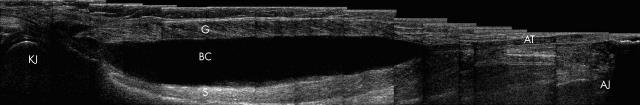
Baker's cyst (photomontage). Photomontage longitudinal image of the calf: Hypoechoic effusion is seen immediately posterior to the knee joint (KJ). This initially extends superficially to the medial head of the gastrocnemius (G) and then dissects between the gastrocnemius and the soleus (S) towards the ankle joint (AJ) for the proximal two thirds of the calf, ending proximal to the formation of the Achilles tendon (AT) by the soleus and gastrocnemius.
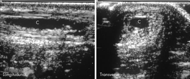
Achilles tendon tear. (A) Longitudinal and (B) transverse images of the calf: at the middle third of the left Achilles tendon there is an anechoic collection (C) lying within the Achilles tendon. A small area of disruption (T) of the deep part of the Achilles tendon fibrillar structure is adjacent to the collection. The findings are consistent with a left Achilles tendon fluid collection resulting from an atraumatic partial Achilles tendon tear (T).
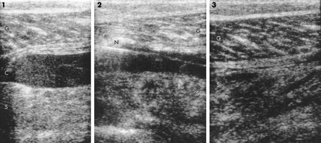
"Tennis Leg". Serial Longitudinal images of the calf: (1) There is a well localised hypoechoic collection (C) interposed between the medial head of the gastrocnemius (G) and the soleus muscle (S), typical of a tear. (2) About 30 ml of blood stained fluid was drained with a 21G needle (N) under ultrasound guidance. (3) This resulted in the collapse of the central hypoechoic fluid collection with restoration of the normal apposition of the gastrocnemius and soleus muscles.
Publication types
MeSH terms
LinkOut - more resources
Full Text Sources
Medical
