. 2003 Dec 10;23(36):11479-88.
doi: 10.1523/JNEUROSCI.23-36-11479.2003.

Effects of neurotoxic and neuroprotective agents on peripheral nerve regeneration assayed by time-lapse imaging in vivo

Affiliations

Effects of neurotoxic and neuroprotective agents on peripheral nerve regeneration assayed by time-lapse imaging in vivo

Y Albert Pan et al. J Neurosci. .

Abstract

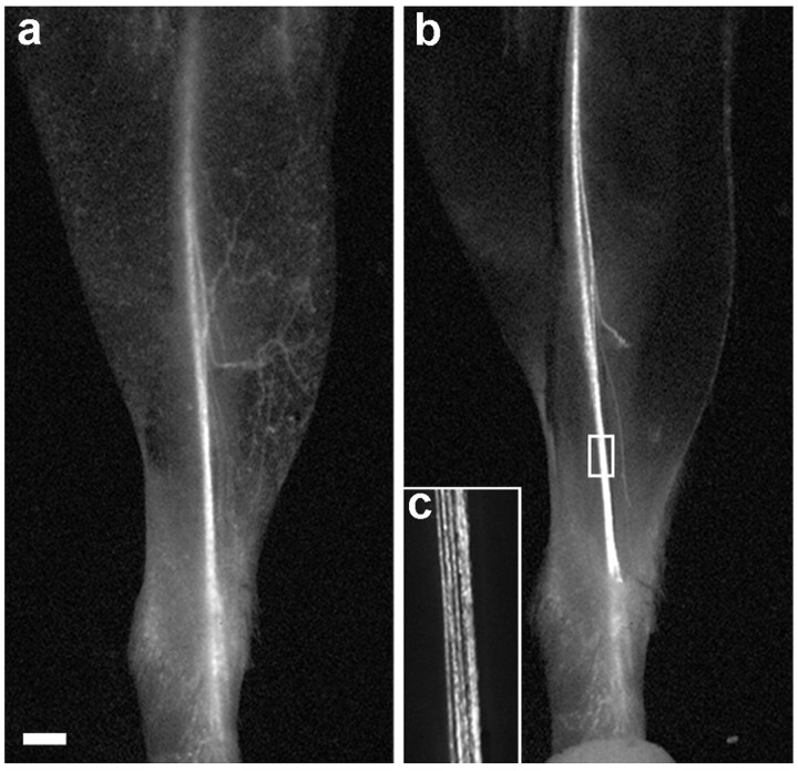
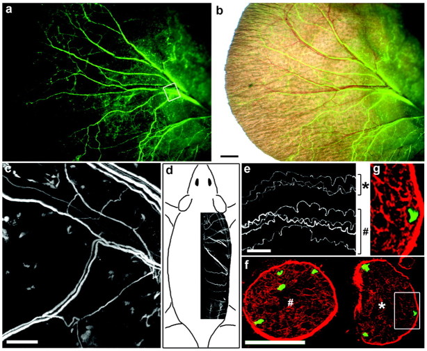
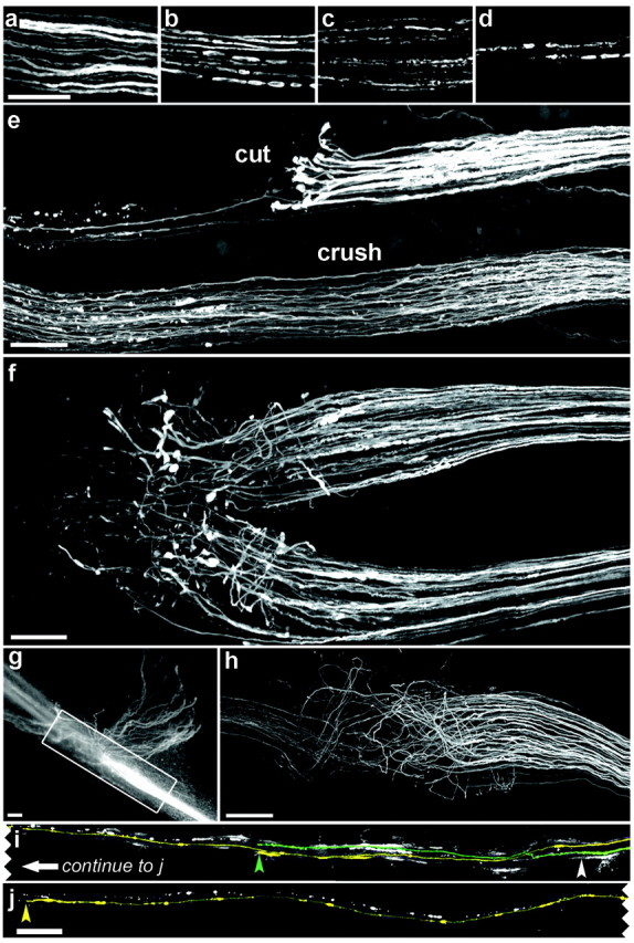
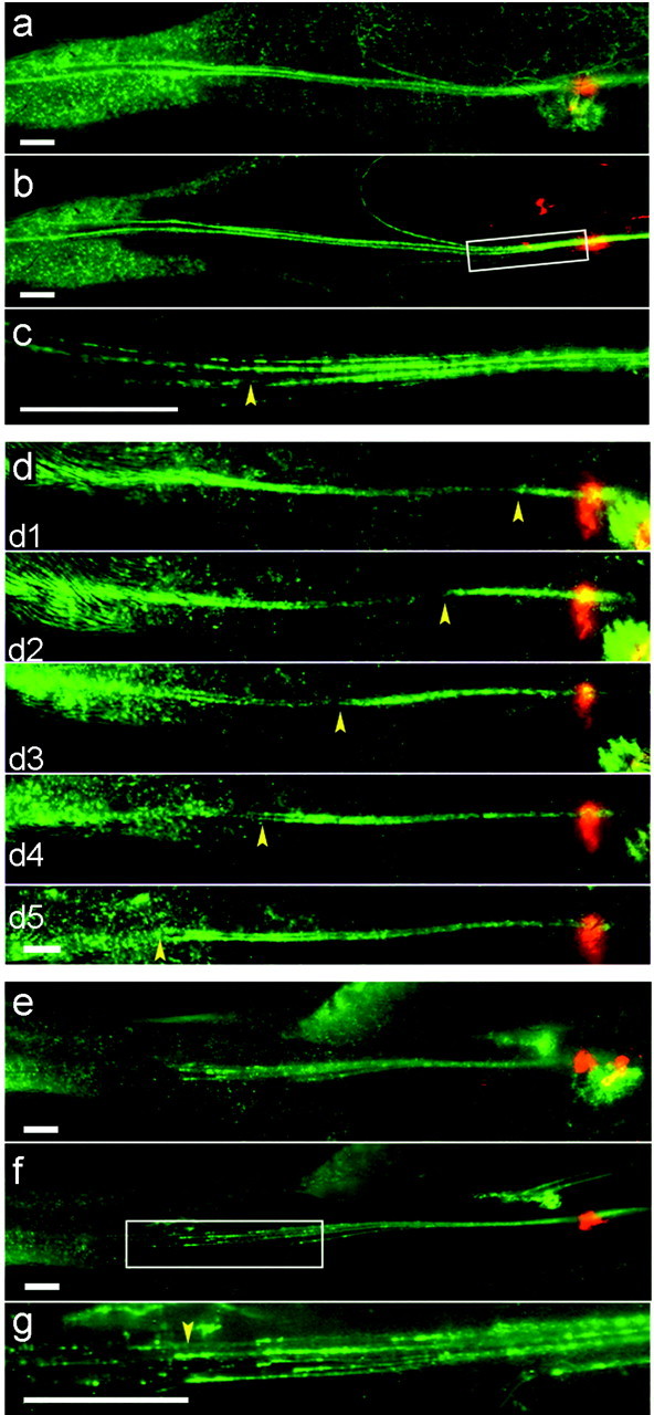
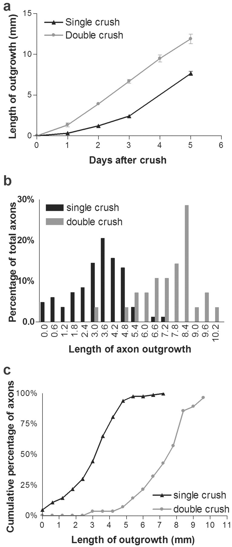
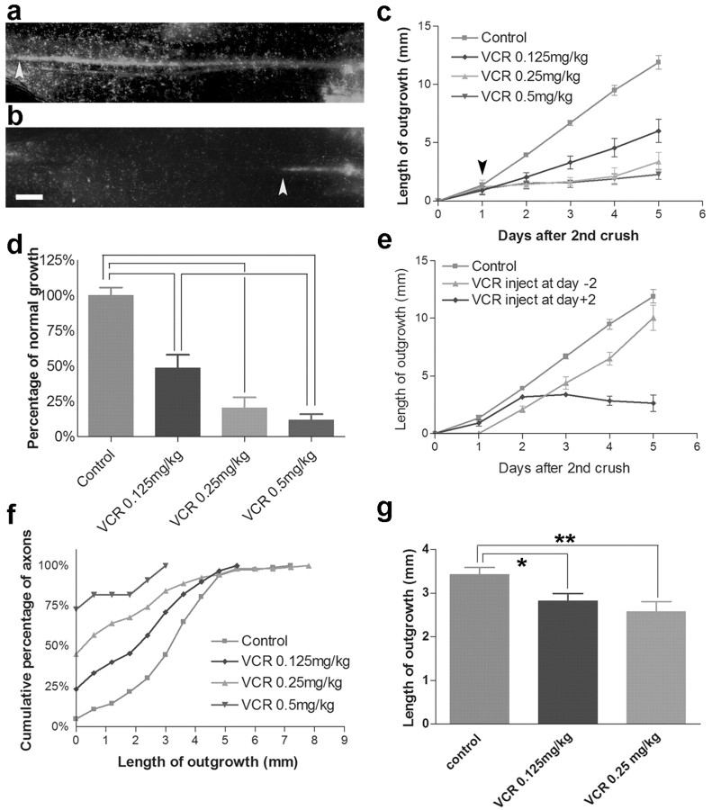
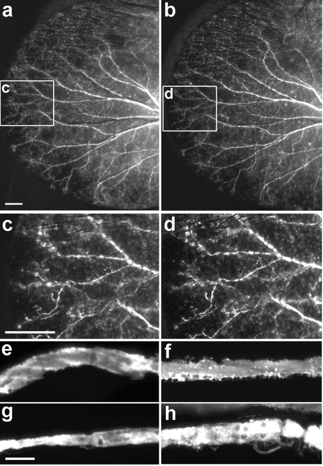
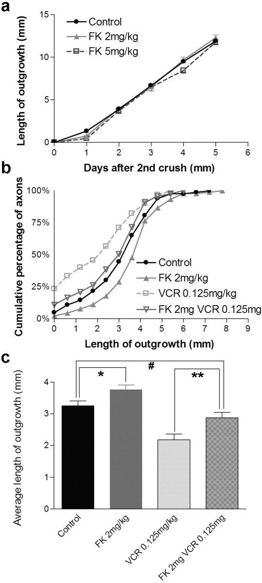
A direct histological assay of axonal regeneration would have many advantages over currently available behavioral, electrophysiological, and radiometric assays. We show that peripheral sensory axons marked with the yellow fluorescent protein in transgenic mice can be viewed transcutaneously in superficial nerves. Degenerating and regenerating axons can be followed in live animals with a dissecting microscope and then, after fixation, studied at high resolution by confocal microscopy. Using this approach, we document differences in regenerative ability after nerve transection, crush injury, and crush injury after a previous "conditioning" lesion. We also show that the chemotherapeutic drug vincristine rapidly but transiently blocks regeneration and that the immunosuppressive drug FK506 modestly enhances regeneration. Moreover, FK506 nearly restores normal regenerative ability in animals treated with submaximal doses of vincristine. Because neuropathy is the major dose-limiting side effect of vincristine, we propose that its efficacy could be enhanced by coadministration of FK506 analogs that are neuroactive but not immunosuppressive.


Figures

Figure 1.
Figure 1.
Transcutaneous imaging of the saphenous nerve. a, The leg of a YFP-H transgenic mouse imaged with a fluorescence dissecting microscope after depilation. The saphenous nerve and some of its cutaneous branches are visible. b, After the image in a was captured, the mouse was killed, the skin was removed, and the leg was imaged again. The boxed area in b is shown at higher magnification in c. Scale bar, 1 mm.
Figure 2.
Figure 2.
Imaging individual axons in the ear (a-c) and dorsal cutaneous nerves (d-g) of YFP-H mice. a, The pinna of the ear of a live mouse. Small bundles of YFP-positive axons are clearly visible. b, Fluorescence image overlaid on a bright-field view. Some but not all axon bundles are apposed to blood vessels. c, After the images in a and b were captured, the animal was killed, the skin was peeled off, and the area boxed in a was imaged again on the confocal microscope to show that individual axons had been visible in the live animal. d, Dorsal cutaneous nerves inserted in a sketch to show their position. e, Confocal image of individual axons in a fixed dorsal cutaneous nerve. f, A region adjacent to that in e was frozen, sectioned, and stained with anti-laminin. The seven axons visible in the whole mount are all seen in the section. Symbols (* and #) mark corresponding nerves in e and f. g, The area boxed in f is shown at higher magnification to demonstrate that the YFP-positive structure occupies a single laminin-rich endoneurial tube and is therefore a single axon. Scale bars: b, 1 mm; c, e, 100 μm; (in f) f, 50 μm, g, 23 μm.
Figure 3.
Figure 3.
Degeneration and regeneration of YFP-positive axons in the saphenous (e-h) and dorsal cutaneous (a-d, i, j) nerves. All panels show confocal images except g, which is from a dissecting microscope. Proximal is to the right in all parts. a-d, Distal stumps 1 d (a), 2 d (b), 3 d (c), or 5 d (d) after transection. YFP-positive axons fragment into ovoids in ∼2 d, some of which persist for >5 d. e, Two branches of the saphenous, 4 d after the top branch had been cut and the bottom had been crushed. Axons thickened after nerve cut and formed club-shaped endings; only a few regenerated into the distal stump (to the left). In contrast, axonal regeneration is advanced after crush injury. f, Six days after the cut, numerous sprouts extend past the site of damage, but they are disordered. g, h, Two weeks after the cut, numerous axons have regenerated, but many of these grow blindly into connective tissue (g), and only a subset grows in the distal stump. i, j, Adjacent portions of the same dorsal cutaneous nerve were imaged 5 d after it was crushed (white arrowhead). There were only two labeled axons in this dorsal cutaneous nerve, both of which have regenerated. The axons are pseudocolored and end at the arrowhead of the corresponding color. White slashes are debris from degenerating YFP-positive axons. Scale bars: a-h, 100 μm; j, 250 μm.
Figure 4.
Figure 4.
Imaging regeneration in live mice after crush injury to the saphenous nerve. Crush sites are indicated by deposits of orange microspheres, and proximal is to the right. Images were captured from a dissecting microscope transcutaneously (a, d, e) or after killing the mice and removing the skin (b, c, f, g). a-c, Three days after a single crush, regeneration is difficult to assess transcutaneously (a). Imaging after removal of skin shows that this is because regenerating axons are intermingled with fluorescent remains of degenerating axons (b, c). The region boxed in b is shown at higher power in c. d, When regeneration is delayed by a double-crush protocol, debris has been cleared by the time regeneration occurs, so the front of regeneration (yellow arrowheads) is easily discerned. d1-d5, 1-5 d after second crush. e-g, The front of fluorescence was viewed transcutaneously in another mouse 5 d after the second of two nerve crushes (e), and then the mouse was killed, the skin was removed, and the same area was imaged again (f). The region boxed in f is shown at higher power in g. The extent of regeneration measured live accurately reflects tips of fastest-regenerating axons (arrowhead). Scale bars, 1 mm.
Figure 5.
Figure 5.
Effect of a conditioning lesion on regeneration after nerve crush. a, Extent of regeneration in the saphenous nerve after a single nerve crush or the second of two crushes. Measurements were made as shown in Figure 4d-g. Points and bars show mean ± SEM (n = 4-8). b, Regeneration of single axons in dorsal cutaneous nerves 5 d after a single nerve crush or the second of two crushes. Measurements were made as shown in Figure 3, i and j (n = 83 axons for single crush and 28 for double crush). c, Cumulative frequency plot of data from b. The difference between curves is significant at p < 0.01 by Kolmogorov-Smirnov test.
Figure 6.
Figure 6.
Inhibition of regeneration by vincristine. a, b, Regeneration in the saphenous 5 d after the second of two nerve crushes in an untreated mouse (a) or a mouse that had received a single injection of 0.5 mg/kg vincristine 4 d previously (b). Arrowheads mark the extent of regeneration. Scale bar, 1 mm. c, d, Dose-dependent inhibition of regeneration by vincristine, measured as in a, b. The arrow in c indicates the time of vincristine injection. Points and bars show mean ± SEM (n = 6-12). Values in d are calculated from the data in c, on the basis of the extension during the 4 d after administration of vincristine. Differences between pairs of bars connected by brackets are significant at p < 0.01 by t test. e, Inhibition of regeneration by vincristine is rapid but transient. Experiments were as in a-c, but the time of vincristine administration (0.25 mg/kg) was either 2 d before (day -2) or 2 d after (day +2) the second crush. f, g, Effects of vincristine on regeneration of single axons in the dorsal cutaneous nerve. The cumulative frequency plot in f shows a dose-dependent increase in the number of axons that fail to regenerate. Lengths of those axons that did regenerate (g) are also decreased by vincristine. The difference between curves in f is significant at p < 0.05 by Kolmogorov-Smirnov test. In g,*p = 0.01; **p = 0.002.
Figure 7.
Figure 7.
A single dose of vincristine does not cause axonal degeneration. a-d, Axons viewed transcutaneously in the pinna of the ear, before and 3 d after administration of vincristine (0.5 mg/kg). The areas boxed in a and b are shown at higher power in c and d. e, f, Confocal images of dorsal cutaneous axons from intact nerves (e, f) or proximal segments of crushed nerves (g, h) in untreated (e, g) or vincristine-treated (0.5 mg/kg) (f, h) mice. Scale bars: a, c, 1 mm; f, 10 μm.
Figure 8.
Figure 8.
FK506 reverses inhibition of regeneration by vincristine. a, Extent of regeneration in the saphenous nerve of mice that received daily doses of FK506. Measurements were made as shown in Figure 4d-g. The points and bars show mean ± SEM (n = 7-10). FK506 has no significant effect on regeneration in this assay, which measured the regenerative rate of the fastest-growing axons. b, Cumulative frequency plot of single axons in dorsal cutaneous nerves from animals that received a single dose of vincristine (VCR), daily administration of FK506, or both. Measurements were made as shown in Figure 3, i and j. n = 83-91 axons per treatment. FK506 modestly speeds regeneration rate. The difference between vincristine and FK plus VCR is significant at p < 0.05 by Kolmogorov-Smirnov test. c, Mean lengths of regenerating axons from data in b. Significance of differences, calculated by t test, were *p = 0.03; **p = 0.005; #p > 0.1.

References

    1. Aley KO, Reichling DB, Levine JD ( 1996) Vincristine hyperalgesia in the rat: a model of painful vincristine neuropathy in humans. Neuroscience 73: 259-265. - PubMed
    1. Apfel SC, Arezzo JC, Lewis ME, Kessler JA ( 1993) The use of insulin-like growth factor I in the prevention of vincristine neuropathy in mice. Ann NY Acad Sci 692: 243-245. - PubMed
    1. Bignami A, Dahl D, Nguyen BT, Crosby CJ ( 1981) The fate of axonal debris in Wallerian degeneration of rat optic and sciatic nerves. Electron microscopy and immunofluorescence studies with neurofilament antisera. J Neuropathol Exp Neurol 40: 537-550. - PubMed
    1. Boyd JG, Gordon T ( 2003) Neurotrophic factors and their receptors in axonal regeneration and functional recovery after peripheral nerve injury. Mol Neurobiol 27: 277-324. - PubMed
    1. Bradley WG, Lassman LP, Pearce GW, Walton JN ( 1970) The neuromyopathy of vincristine in man. Clinical, electrophysiological and pathological studies. J Neurol Sci 10: 107-131. - PubMed

Publication types

MeSH terms

LinkOut - more resources