. 2003 Dec 23;100(26):15566-71.
doi: 10.1073/pnas.2536695100. Epub 2003 Dec 12.

The kinesin KIF17b and RNA-binding protein TB-RBP transport specific cAMP-responsive element modulator-regulated mRNAs in male germ cells

Affiliations

The kinesin KIF17b and RNA-binding protein TB-RBP transport specific cAMP-responsive element modulator-regulated mRNAs in male germ cells

Vargheese Chennathukuzhi et al. Proc Natl Acad Sci U S A. .

Abstract

Testis brain RNA-binding protein (TB-RBP), the mouse orthologue of the human protein Translin, is a widely expressed and highly conserved protein with proposed functions in chromosomal translocations, mitotic cell division, and mRNA transport, stabilization, and storage. Targeted inactivation of TB-RBP leads to abnormalities in fertility and behavior. A testis-enriched kinesin KIF17b coimmunoprecipitates with TB-RBP in a RNA-protein complex containing specific cAMP-responsive element modulator (CREM)-regulated mRNAs. The specificity of this interaction is confirmed by in vivo RNA-protein crosslinking and transfections of hippocampal neurons. Combining in situ hybridization and immunohistochemistry at the electron microscope level, a temporally sequential dissociation of KIF17b and TB-RBP from specific mRNAs is detected with TB-RBP release coincident with the time of mRNA translation, indicating a separation of the processes of transport and translation. We conclude that KIF17b serves as a molecular motor component of a TB-RBP-mouse ribonucleoprotein complex transporting a group of specific CREM-regulated mRNAs in mammalian male postmeiotic germ cells. Because KIF17b has been reported to control CREM-dependent transcription in male germ cells by regulating the intracellular location of the transcriptional coactivator activator of CREM in testis, this indicates that one kinesin links the processes of transcription and transport of specific mRNAs in mammalian male germ cells.


Figures

Fig. 1.
Fig. 1.
(A) Coimmunoprecipitation of KIF17b and TB-RBP from mouse testis extracts. A silver-stained SDS/polyacrylamide gel shows total extracts from brain (lane 1) and testis (lane 2), protein A-agarose beads preclearing controls of extracts from brain (lane 3) and testis (lane 4), and immunoprecipitated cytoplasmic S100 extracts from brain (lane 5) and testis (lane 6) using affinity-purified anti-TB-RBP. Excised protein bands were sequenced by MALDI-TOF mass spectrometry. M, marker proteins. (B) KIF17b expression is specific to postmeiotic male germ cells. Shown is a Northern blot of RNA (10 μg) from brain (lane 1), testis (lane 2), mixed germ cells (lane 3), pachytene spermatocytes (lane 4), round spermatids (lane 5), and elongating spermatids (lane 6). Robust expression of KIF17b mRNA is seen in the postmeiotic male germ cells. (C) Affinity-purified anti-KIF17b recognizes TB-RBP-associated KIF17b from testis extracts. A Western blot shows protein A-agarose beads preclearing of extracts from brain (lane 1) and testis (lane 2), brain S100 cytoplasmic extract (lane 3), anti-TB-RBP immunoprecipitation of extracts from brain (lane 4) and testis (lane 5), and the enrichment of KIF17b in the TB-RBP immunoprecipitate (lane 6). (D) Anti-KIF17b coimmunoprecipitates TB-RBP from testis extracts. Western blot of extract immunoprecipitated with anti-KIF17b antibodies shows TB-RBP in the immunoprecipitate. Lane 1, protein A-agarose bead preclearing; lane 2, anti-KIF17b immunoprecipitate.
Fig. 3.
Fig. 3.
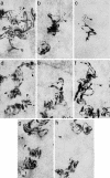
Colocalization of TB-RBP, KIF17b, and mRNAs by in situ hybridization and immunoradioautography. Shown are TB-RBP (15-nm gold particles, solid arrowheads) and KIF17b (10-nm gold particles, open arrowheads) in mouse seminiferous tubules hybridized in situ with antisense RNA probes (large filamentous silver grains) encoding AKAP4 (a, b, d, e, and g), protamine 2 (c and f), or PGK2 (h) In early (a, step 2) and late (b, step 7) round spermatids, AKAP4 silver grains are in close association with KIF17b and TB-RBP (10- and 15-nm colloidal gold particles (a, step 2). Protamine 2-generated silver grains are also in close association with KIF17b and TB-RBP in round spermatids (c, step 7) whereas PGK2-generated silver grains are not (h, step 7). During spermatid elongation, the KIF17b gold particles gradually dissociate from AKAP4 mRNAs (d, step 9, and e, step 11) and from the protamine mRNAs (f, step 11). In late step spermatids TB-RBP also dissociates from the AKAP4 mRNAs (g, step 15) and from the protamine 2 mRNAs (data not shown). (Magnification: ×100,000.)
Fig. 2.
Fig. 2.
Immunoprecipitation assay showing TB-RBP binding to specific germ cell mRNAs. RNA protein complexes immunoprecipitated after in vivo formaldehyde crosslinking in trypsinized testicular cells were analyzed by RT-PCR (35 cycles of amplification) using specific primers for each mRNA. Lane 1, total RNA from TB-RBP–/– testes; lane 2, preclearing of crosslinked extracts; lane 3, immunoprecipitate from wild-type testes with anti-TB-RBP; lane 4, immunoprecipitate from TB-RBP–/– testes with anti-TB-RBP; lane 5, immunoprecipitate from wild-type testes with anti-KIF17b; lane 6, immunoprecipitate from TB-RBP–/– testes with anti-KIF17b. RNAs were resolved on agarose gels and visualized with ethidium bromide staining.
Fig. 4.
Fig. 4.
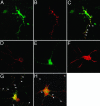
Transfected KIF17b colocalizes with TB-RBP and Y and H element containing RNA in rat hippocampal neurons. Plasmid constructs containing KIF17b and TB-RBP cDNAs cloned in frame in pEGFPc2 and pDsRed1 vectors, respectively, were transfected into cultured rat embryonic hippocampal neurons with Nupherin-neuron and FuGENE 6 reagent. (A) GFP–KIF17b fusion protein. (B) TB-RBP–DsRed fusion protein. (C) Merged image. Arrowheads indicate the colocalization of KIF17b and TB-RBP in the dendritic processes. (DH) Transfected Y and H element RNA colocalizes with TB-RBP and KIF17b. Cells expressing GFP–TB-RBP or GFP–KIF17b were retransfected 36 h later with Alexa Fluor 546 RNA probe and show colocalization (arrows) of RNA with GFP–TB-RBP (G) and GFP–KIF17b (H). Control single transfections with TB-RBP–DsRed (D), GFP–KIF17b (E), and Alexa Fluor 546 (F) riboprobes. Dual-channel confocal imaging was performed within 30 min of transfection of the riboprobe.

Similar articles

Cited by

References

    1. Braun, R. E., Behringer, R. R., Peschon, J. J., Brinster, R. L. & Palmiter, R. D. (1989) Nature 337, 373–376. - PubMed
    1. Caldwell, K. A. & Handel, M. A. (1991) Proc. Natl. Acad. Sci. USA 88, 2407–2411. - PMC - PubMed
    1. Zou, Y., Millette, C. F. & Sperry, A. O. (2002) Biol. Reprod. 66, 843–855. - PubMed
    1. Aoki, K., Suzuki, K., Sugano, T., Tasaka, T., Nakahara, K., Kuge, O., Omori, A. & Kasai, M. (1995) Nat. Genet. 10, 167–174. - PubMed
    1. Morales, C. R., Lefrancois, S., Chennathukuzhi, V., El-Alfy, M., Wu, X., Yang, J., Gerton, G. L. & Hecht, N. B. (2002) Dev. Biol. 246, 480–494. - PubMed

Publication types

MeSH terms

Associated data