. 2003 Dec 23;100(26):15818-23.
doi: 10.1073/pnas.2636938100. Epub 2003 Dec 12.

Antigen-driven effector CD8 T cell function regulated by T-bet

Affiliations

Antigen-driven effector CD8 T cell function regulated by T-bet

Brandon M Sullivan et al. Proc Natl Acad Sci U S A. .

Abstract

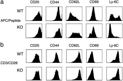
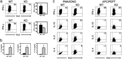
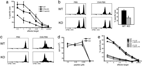
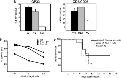
Type 1 immunity relies on the differentiation of two major subsets of T lymphocytes, the CD4+ T helper (Th) cell and the CD8+ cytotoxic T cell, that direct inflammatory and cytotoxic responses essential for the destruction of intracellular and extracellular pathogens. In contrast to CD4 cells, little is known about transcription factors that control the transition from the CD8 naïve to effector cell stage. Here, we report that the transcription factor T-bet, known to regulate Th cell differentiation, also controls the generation of the CD8+ cytotoxic effector cell. Antigen-driven generation of effector CD8+ cells was impaired in OT-I T cell receptor transgenic mice lacking T-bet, resulting in diminished cytotoxicity and a marked shift in cytokine secretion profiles. Furthermore, mice lacking T-bet responded poorly to infection with lymphocytic choriomeningitis virus. T-bet is a key player in the generation of type 1 immunity, in both Th and T cytotoxic cells.


Figures

Fig. 1.
Fig. 1.
T-bet-deficient CD8+ T cells fail to acquire an effector phenotype after antigenic stimulation. OT-I CD8 T cells were isolated by magnetic bead purification from the lymph nodes of T-bet-/- and WT OT-I TCR transgenic mice. Cells were stimulated with irradiated C57BL/6 splenocytes and peptide (1 μM) (a) or plate-bound anti-CD3 and anti-CD28 with recombinant human IL-2 (b) for 72 h, then expanded. Populations were stained with PE (CD25, CD44, CD62L)- and FITC (CD69, Ly6C)-conjugated antibodies and analyzed by FACS. KO, knockout.
Fig. 2.
Fig. 2.
Diminished IFN-γ, but increased IL-2 and Tc2 cytokine production by antigen-stimulated T-bet-deficient CD8 T cells. T-bet-/- and WT OT-I CD8 cells were stimulated with APC/peptide (a Upper, b, and c) or anti-CD3, anti-CD28, and recombinant human IL-2 (a Lower), as in Fig. 1. (a) Defective IFN-γ production by T-bet-/- OT-I CD8 cells only under antigen-specific stimulation conditions. Cells were restimulated for 24 h by either APC/peptide (Upper Right) or plate-bound anti-CD3 (Lower Right), and secreted IFN-γ was measured by ELISA. Intracellular IFN-γ staining was performed after stimulation with either APC/peptide (Upper) for 18 h or PMA/ionomycin (Lower)for 5 h. (b) Elevated levels of IL-2 and Tc2 cytokines in T-bet-deficient CD8 cells. IL2, IL-4, and IL10 production was measured by ELISA of supernatants taken after a 24-h secondary APC/peptide stimulation. (c) The altered cytokine profile of antigen-stimulated T-bet-/- OT-I CD8 cells is independent of secondary stimulation conditions. After an initial 72-h APC/peptide stimulation and expansion, cells were restimulated for intracellular cytokine analysis by either PMA/ionomycin (Left) or APC/peptide (Right). Dot plot values represent the percentage of Vα2+ cells that stained positive for the indicated cytokine. KO, knockout.
Fig. 3.
Fig. 3.
Defective CTL effector function in T-bet-deficient OT-I CD8 cells. (a and e) WT, T-bet-/-, and IFN-γ-/- OT-I CD8 cells were stimulated with APC/peptide for 72 h, harvested, and then incubated for 6 h with 51Cr-labeled, peptide-pulsed EL4 targets. Data presented represent triplicate reactions at each effector/target ratio from one experiment (n = 5). (b) Lymph node T-bet-/- and WT OT-I CD8 cells were enriched by combined CD4 and CD19 negative selection and transferred to C57BL/6 hosts i.v. Recipients were immunized by s.c. injection with either PBS (control) or ovalbumin (experimental). After 6 days, equal numbers of peptide-pulsed CFSE-hi targets and unpulsed CFSE-lo control splenocytes were injected i.v. to each host. The draining inguinal nodes were processed 10 h later, and specific lysis was assessed by FACS. Histograms represent propidium iodide-negative, CFSE-positive acquisitions from one mouse per group and are representative of two independent experiments. Specific lysis was also quantified and graphed as the mean ± SEM of both experiments, totaling six recipient mice per group. (c and d) T-bet-/- OT-I CD8 cells proliferate comparably to WT controls. (c) CFSE-labeled T-bet-/- and WT OT-I, CD8 cells were labeled and transferred to WT C75BL/6 hosts. Recipients were immunized as described above (b). The draining nodes were harvested 2 days after ovalbumin immunization, stained with CD8-CyC and Vα2-PE antibodies, and analyzed by FACS. Histograms represent CFSE intensity of CD8+, Vα2+ gated populations. (d) OT-I, CD8 cells were stimulated with splenocytes and peptide at the indicated concentrations for 72 h. Reactions were performed in triplicate and pulsed with [3H]thymidine for the final 24 h of stimulation. (e) Diminished CTL capacity cannot be attributed to the imbalance in cytokine production. Purified OT-I, CD8 cells were stimulated as described in the presence of recombinant murine IL-10 (10 μg/ml), recombinant murine IFN-γ (10 ng/ml), anti-IL10 (10 μg/ml), or anti-IFN-γ (75 μg/ml) for 72 h, followed by CTL analysis. KO, knockout.
Fig. 4.
Fig. 4.
T-bet-deficient mice fail to generate substantial viral-specific CTL activity and exhibit inadequate protection against LCMV infection. (a) Spleen cells of LCMV-infected mice were cultured with peptide, GP33, or plate-bound anti-CD3/CD28. IFN-γ production was assessed by intracellular staining and presented as percent IFN-γ+ of gated CD8+ populations. Data represent the mean ± SEM of two to three mice per group. (b) LCMV-infected T-bet-/-, T-bet+/-, and T-bet+/+ spleen cells were cultured for 6 days with LCMV-infected peritoneal exudate cells. CTL activity was quantified as 51Cr release from LCMV-infected MC57 cells at the indicated effector/target ratios. (c) T-bet-/- and T-bet+/- or T-bet+/+ mice were vaccinated with pCMV-NP, 7 and 14 days before intracranial LCMV challenge. naïve, unimmunized mice served as controls for lack of protection against infection. KO, knockout; HET, heterozygote.

References

    1. Kaech, S. M., Wherry, E. J. & Ahmed, R. (2002) Nat. Rev. Immunol. 2, 251-262. - PubMed
    1. Harty, J. T. & Badovinac, V. P. (2002) Curr. Opin. Immunol. 14, 360-365. - PubMed
    1. Wong, P. & Pamer, E. G. (2003) Annu. Rev. Immunol. 21, 29-70. - PubMed
    1. Carter, L. L. & Murphy, K. M. (1999) J. Exp. Med. 189, 1355-1360. - PMC - PubMed
    1. Nguyen, K. B., Watford, W. T., Salomon, R., Hofmann, S. R., Pien, G. C., Morinobu, A., Gadina, M., O'Shea, J. J. & Biron, C. A. (2002) Science 297, 2063-2066. - PubMed

Publication types

MeSH terms

Substances

LinkOut - more resources