. 2003 Dec 23;100(26):15806-11.
doi: 10.1073/pnas.2636971100. Epub 2003 Dec 12.

Diversity of regulatory CD4+T cells controlling distinct organ-specific autoimmune diseases

Affiliations

Diversity of regulatory CD4+T cells controlling distinct organ-specific autoimmune diseases

Marie-Alexandra Alyanakian et al. Proc Natl Acad Sci U S A. .

Erratum in

  • Proc Natl Acad Sci U S A. 2004 Mar 23;101(12):4331

Abstract

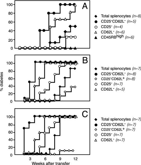
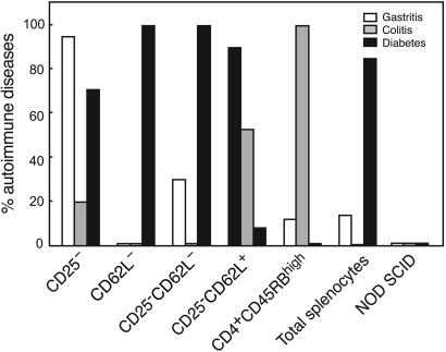
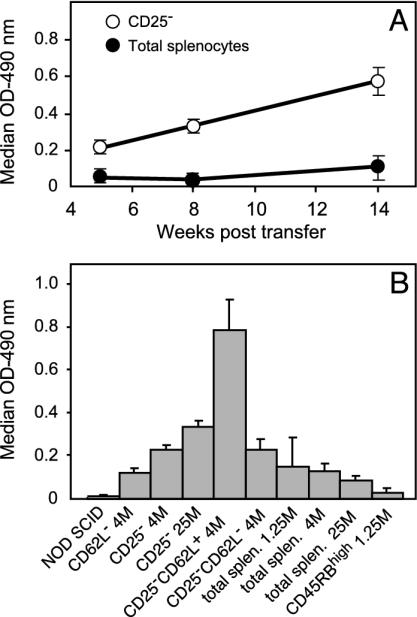
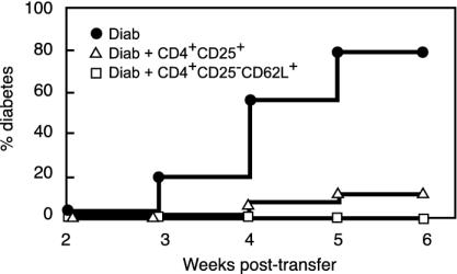
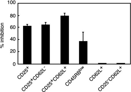
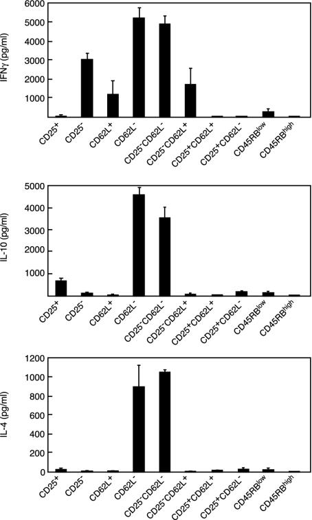
Depletion of selected regulatory CD4+ T cell subsets induces the spontaneous onset of various immune or autoimmune disorders. It is not clear, however, whether a given subset, notably CD4+CD25+ regulatory T cells, protects from a wide spectrum of immune disorders, or whether specialized subsets of regulatory T cells control each given disease or group of diseases. We report here, using diabetes prone nonobese diabetic (NOD) mice, that depending on the regulatory T cells that are depleted, i.e., CD25+, CD62L+, or CD45RB(low), distinct immune diseases appear after transfer into NOD severe combined immunodeficiency (SCID) recipients. Thus, reconstitution of NOD SCID mice with CD25- T cells induces major gastritis and late-onset diabetes, but no or mild colitis. Reconstitution with CD62L- T cells induces fulminant diabetes with no colitis or gastritis. Reconstitution with CD45RB(high) T cells induces major colitis with wasting disease and no or very moderate gastritis and diabetes. Major differences among the three regulatory T cell subsets are also seen in vitro. The bulk of suppressor cells inhibiting the proliferation of CD4+CD25- T cells in coculture is concentrated within the CD25+ but not the CD62L+ or CD45RB(low) T cell subsets. Similarly, cytokine production patterns are significantly different for each regulatory T cell subset. Collectively, these data point to the diversity and organ selectivity of regulatory T cells controlling distinct autoimmune diseases whatever the underlying mechanisms.


Figures

Fig. 1.
Fig. 1.
Diversity of regulatory CD4+ T cells controlling autoimmune diabetes. Results of a representative experiment show diabetes incidence in NOD SCID mice reconstituted with total spleen cells or spleen cells depleted in distinct regulatory T cell subsets from 10-wk-old NOD mice. Independent from the cell population analyzed, diabetes incidence correlated with the number of transferred cells, i.e., 1.25 × 10⁶ (A), 4 × 10⁶ (B), and 7 × 10⁶ (C). The data highlight three main points. First, CD25-CD62L- T cells are significantly more effective in transferring diabetes than total spleen cells [P < 0.01 (A), P < 0.0002 (B), and P < 0.0001 (C)]. Second, purified CD25-CD62L- T cells triggered extremely severe diabetes at an incidence significantly higher than that observed in recipients of CD25-CD62L+ T cells [P < 0.0001 (B) and P < 0.0001 (C)]. Third, CD4+CD45RBhigh T cells that are very effective at transferring colitis were not diabetogenic at all.
Fig. 2.
Fig. 2.
Gastritis, colitis, and diabetes in NOD SCID mice reconstituted with spleen cells depleted of distinct regulatory T cell subsets. Incidence of overt diabetes and histological gastritis and colitis in a total of 194 NOD SCID mice reconstituted with various T cell subsets is shown. The major finding was that CD25-CD62L+, CD4+CD45RBhigh, and CD4+CD62L- T cells were unique in their capacity to trigger almost exclusively one disease, namely gastritis, colitis, or diabetes, respectively. Intermediate patterns, both in terms of incidence and severity (see also Table 1), were observed for the other purified subsets.
Fig. 3.
Fig. 3.
Anti-H+/K+ ATPase autoantibody serum levels. Specific autoantibody levels were determined by ELISA. (A) Kinetics of anti-H+/K+ ATPase autoantibody production in mice reconstituted with 4 × 10⁶ of either total spleen cells (•) or purified CD25- T cells (○). (B) Antibody levels at 12 wk posttransfer in the various groups of recipients analyzed. The x axis details the phenotype and the number (M, millions of cells) for each transferred lymphocyte subset. Mean antibody levels detected in recipients injected with CD25-CD62L+ T cells were significantly higher than those observed in all other groups (P < 0.001).
Fig. 4.
Fig. 4.
Spleen CD4+CD25+ and CD4+CD25-CD62L+ T cells from prediabetic NOD mice are protective in vivo. Diabetes was monitored in NOD SCID recipients injected with diabetogenic cells alone (Diab) or with mixtures of diabetogenic cells and 1 × 10⁶ CD4+CD25+ or CD4+CD25-CD62L+ purified T cells. In the two latter groups, significant diabetes protection was observed (P < 0.0001 for CD4+CD25+ and CD4+CD25-CD62L+). Shown are combined data of six to seven independent experiments.
Fig. 5.
Fig. 5.
In vitro suppression of CD4+CD25- T cell proliferation by distinct regulatory T cell subsets. Data collected from different experiments are expressed as percent inhibition of proliferation. Results show that cells expressing the CD25 marker are the most effective suppressors in vitro. In great distinction, no suppression was exhibited by T cell subsets expressing CD62L but no CD25.
Fig. 6.
Fig. 6.
Cytokine production by selected T cell subsets after CD3 antibody-induced stimulation. Results show that the cytokine-producing ability greatly differed among the distinct subsets analyzed. Mean values observed with CD62L- and CD25-CD62L- T cells were significantly higher for the three cytokines analyzed than those observed for all other T cell subsets tested (P < 0.0001).

References

    1. Shevach, E. M. (2002) Nat. Rev. Immunol. 2, 389-400. - PubMed
    1. Bach, J. F. (2003) Nat. Rev. Immunol. 3, 189-198. - PubMed
    1. Asano, M., Toda, M., Sakaguchi, N. & Sakaguchi, S. (1996) J. Exp. Med. 184, 387-396. - PMC - PubMed
    1. Powrie, F., Leach, M. W., Mauze, S., Caddle, L. B. & Coffman, R. L. (1993) Int. Immunol. 5, 1461-1471. - PubMed
    1. Herbelin, A., Gombert, J. M., Lepault, F., Bach, J. F. & Chatenoud, L. (1998) J. Immunol. 161, 2620-2628. - PubMed

Publication types

MeSH terms