. 2004 Jan;24(1):84-95.
doi: 10.1128/MCB.24.1.84-95.2004.

Specific and covalent targeting of conjugating and deconjugating enzymes of ubiquitin-like proteins

Affiliations

Specific and covalent targeting of conjugating and deconjugating enzymes of ubiquitin-like proteins

Joris Hemelaar et al. Mol Cell Biol. 2004 Jan.

Abstract

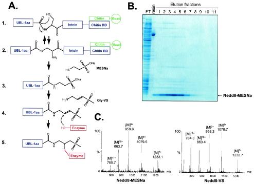
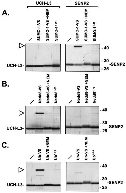
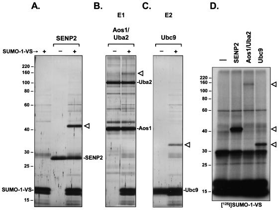
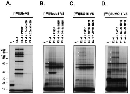
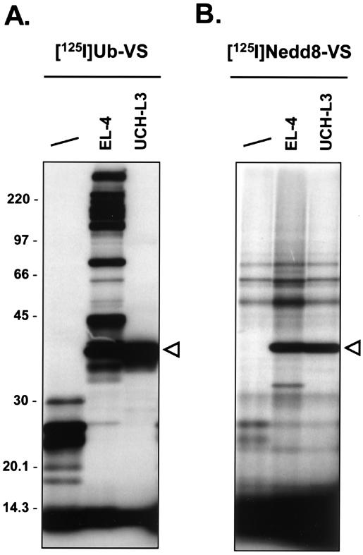
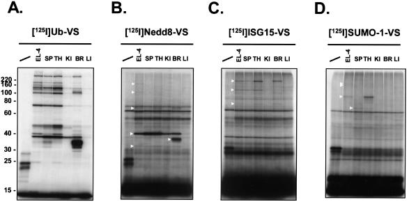
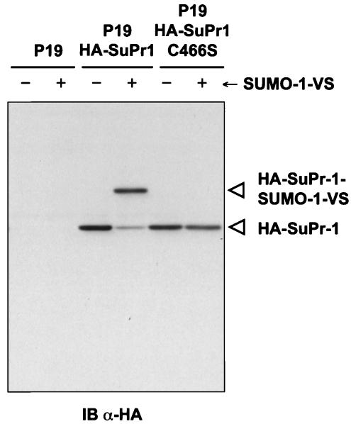
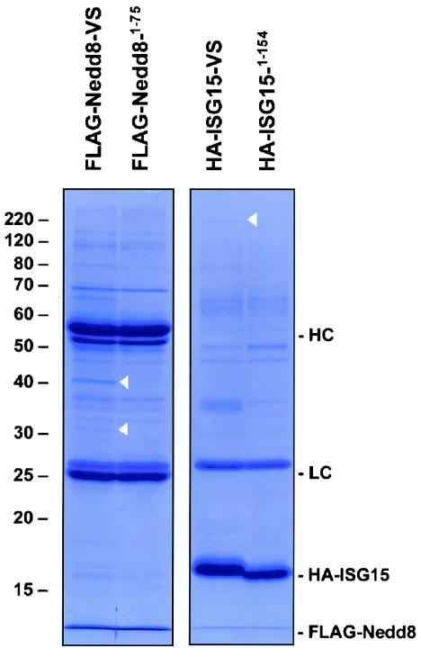
Modification of proteins by ubiquitin (Ub)-like proteins (UBLs) plays an important role in many cellular processes, including cell cycle progression, nuclear transport, and autophagy. Protein modification occurs via UBL-conjugating and -deconjugating enzymes, which presumably exert a regulatory function by determining the conjugation status of the substrate proteins. To target and identify UBL-modifying enzymes, we produced Nedd8, ISG15, and SUMO-1 in Escherichia coli and equipped them with a C-terminal electrophilic trap (vinyl sulfone [VS]) via an intein-based method. These C-terminally modified UBL probes reacted with purified UBL-activating (E1), -conjugating (E2), and -deconjugating enzymes in a covalent fashion. Modified UBLs were radioiodinated and incubated with cell lysates prepared from mouse cell lines and tissues to allow visualization of polypeptides reactive with individual UBL probes. The cell type- and tissue-specific labeling patterns observed for the UBL probes reflect distinct expression profiles of active enzymes, indicating tissue-specific functions of UBLs. We identify Ub C-terminal hydrolase L1 (UCH-L1) and DEN1/NEDP1/SENP8, in addition to UCH-L3, as proteases with specificity for Nedd8. The Ub-specific protease isopeptidase T/USP5 is shown to react with ISG15-VS. Furthermore, we demonstrate that the desumoylation enzyme SuPr-1 can be modified by SUMO-1-VS, a modification that is dependent on the SuPr-1 active-site cysteine. The UBL probes described here will be valuable tools for the further characterization of the enzymatic pathways that govern modification by UBLs.


Figures

FIG. 1.
FIG. 1.
Synthesis of UBLs with a C-terminal VS. (A) Reaction scheme for UBL-VS synthesis. Step 1, the processed form of a UBL, minus the C-terminal amino acid (−1aa), is expressed as a fusion protein with an intein and a CBD. Soluble fusion protein binds to a chitin affinity column. Step 2, spontaneous N-S acyl rearrangement resulting in an intermediate in which the peptide bond is replaced by a thioester linkage. Step 3, the UBL is released from the column by a transthioesterification reaction induced by incubation with MESNa sodium salt, resulting in the UBL-MESNa product. Step 4, the MESNa group is replaced by glycine-VS in a chemical ligation reaction, producing UBL-VS. Step 5, nucleophilic active-site residues of enzymes can covalently react with the VS group. (B) Nedd8-MESNa purification. FT, flowthrough after loading of lysate on chitin column; resin, chitin resin after Nedd8-MESNa elution. Eluted Nedd8-MESNa fractions were collected after overnight on-column cleavage induced by MESNa. Samples were prepared in SDS sample buffer without β-mercaptoethanol. (C) LC-ESI-MS analysis of Nedd8-MESNa and Nedd8-VS conversion product. The indicated multicharged species of Nedd8-MESNa correspond to a molecular weight of 8,627.6 ± 2.3, in agreement with a predicted molecular weight of 8,628.1. For Nedd8-VS, multicharged species correspond to a molecular weight of 8,620.8 ± 2.5, in agreement with a predicted molecular weight of 8,621.1.
FIG. 2.
FIG. 2.
UBL-VSs react specifically with cognate deconjugating enzymes. (A) Recombinant, purified UCH-L3 enzyme (left panel) and the catalytic domain of SENP2 (right panel) were incubated for 1 h at 37°C with SUMO-1 derivatives. No SUMO-1 probe was added to the first sample in each panel. Where indicated, enzymes were pretreated with 10 mM NEM prior to addition of SUMO-1-VS. SUMO1-96 lacks the C-terminal glycine and VS moiety and therefore is not reactive. The reactions were terminated by addition of SDS sample buffer containing β-mercaptoethanol and boiling for 5 min. Polypeptides were resolved by SDS-11% PAGE and visualized by silver staining. The positions of molecular mass markers (in kilodaltons) are indicated on the right. The positions of UCH-L3 and the catalytic domain of SENP2 are indicated. The triangle marks the position of the SENP2-SUMO-1-VS adduct. (B) Same as panel A but with the Nedd8 derivatives Nedd8-VS and Nedd81-75. Nedd81-75 lacks the C-terminal glycine and VS group. The triangle marks the position of the UCH-L3-Nedd8-VS adduct. (C) Same as panel A but with the Ub derivatives Ub-VS and Ub1-75. Ub1-75 lacks the C-terminal glycine and VS group. The triangle marks the position of the UCH-L3-Ub-VS adduct.
FIG. 3.
FIG. 3.
SUMO-1-VS covalently modifies its cognate E1 and E2 enzymes in vitro. (A) Recombinant, purified SENP2 catalytic domain was incubated for 2 h at 37°C with (+) or without (−) SUMO-1-VS. SUMO-1-VS alone is shown in the first lane. The reactions were terminated as described for Fig. 2. Polypeptides were resolved by SDS-11% PAGE and visualized by silver staining. The triangle marks the position of the SENP2-SUMO-1-VS adduct. The positions of molecular mass markers (in kilodaltons) are indicated on the left. (B) Recombinant, purified SUMO-1-specific E1 activating enzyme, composed of the Aos1/Uba2 heterodimer, was incubated for 2 h at 37°C with (+) or without (−) SUMO-1-VS. Samples were subsequently processed as described for panel A. The triangle marks the position of the Uba2-SUMO-1-VS adduct. (C) Recombinant, purified SUMO-1-specific E2 conjugating enzyme, Ubc9, was incubated for 2 h at 37°C with (+) or without (−) SUMO-1-VS. Samples were subsequently processed as described for panel A. The triangle marks the position of the Ubc9-SUMO-1-VS adduct. (D) 125I-SUMO-1-VS (106 cpm) was incubated for 1 h at 37°C with either no enzyme (first lane) or 12.5 pmol of SENP2, Aos1/Uba2, or Ubc9. Polypeptides were resolved by SDS-11% PAGE and visualized by autoradiography. The triangles mark the positions of the observed SUMO-1-VS adducts (top to bottom) Uba2-SUMO-1-VS, SENP2-SUMO-1-VS, and Ubc9-SUMO-1-VS.
FIG. 4.
FIG. 4.
UBL-VSs label distinct sets of proteins in EL-4 lysates. VS derivatives of the indicated UBLs were radiolabeled with Na125I and incubated with EL-4 cell lysates. Per sample, 5 × 10⁵ cpm of 125I-labeled probe and 40 μg of EL-4 lysate were used. First lane in each panel, no lysate added (UBL-VS probe only). EL-4 lysate was pretreated with 1 mM PMSF or with 10 or 20 mM NEM prior to addition of the UBL-VS probes. The reactions were terminated as described for Fig. 2. Polypeptides were resolved by SDS-11% PAGE and visualized by autoradiography. Triangles mark the positions of observed UBL-VS-protein adducts. The radiolabeled UBL-VS probes used were 125I-Ub-VS (A), 125I-Nedd8-VS (B), 125I-UCRP-VS (C), and 125I-SUMO-1-VS (D). The positions of molecular mass markers (in kilodaltons) are indicated on the left.
FIG. 5.
FIG. 5.
UCH-L3 labeling by 125I-Ub-VS and 125I-Nedd8-VS. 125I-Ub-VS (2.5 × 10⁵ cpm) (A) or 125I-Nedd8-VS (2.5 × 10⁵ cpm) (B) was incubated for 1 h at 37°C with lysate buffer (first lane), 20 μg of EL-4 lysate, or 150 ng of recombinant, purified UCH-L3. The reactions were terminated as described for Fig. 2. Polypeptides were resolved by SDS-11% PAGE and visualized by autoradiography. The triangles mark the UCH-L3-Ub-VS (left) and UCH-L3-Nedd8-VS (right) adducts. The positions of molecular mass markers (in kilodaltons) are indicated on the left.
FIG. 6.
FIG. 6.
UBL-VSs exhibit differential labeling patterns in mouse tissue extracts. Lysates from single-cell suspensions of the indicated mouse tissues were prepared and incubated for 1 h at 37°C with radiolabeled UBL-VSs. Per reaction, 5 × 10⁵ cpm of 125I-UBL-VS and 20 μg of EL-4 or tissue lysate were used. The reactions were terminated as described for Fig. 2. Polypeptides were resolved by SDS-10% PAGE and visualized by autoradiography. First lane in each panel, no lysate added (UBL-VS probe only). SP, spleen; TH, thymus; KI, kidney; BR, brain; LI, liver. Triangles mark the positions of observed UBL-VS-protein adducts. The radiolabeled UBL-VS probes used were125I-Ub-VS (A), 125I-Nedd8-VS (B), 125I-UCRP-VS (C), and 125I-SUMO-1-VS (D). The positions of molecular mass markers (in kilodaltons) are indicated on the left.
FIG. 7.
FIG. 7.
SUMO-1 modifies SuPr-1 via its active-site cysteine. P19 cells were transiently transfected with a vector expressing HA-SuPr-1 or the catalytically inactive mutant HA-SuPr-1C466S, cells were harvested, and nuclear extracts were prepared. Nuclear extracts (25 μg) were incubated for 1 h at 37°C without (−) or with (+) SUMO-1-VS. The reactions were terminated by addition of SDS sample buffer containing β-mercaptoethanol and boiling for 5 min. Polypeptides were resolved by SDS-10% PAGE and immunoblotted (IB) with an anti-HA (α-HA) antibody.
FIG. 8.
FIG. 8.
Isolation of polypeptides reactive with FLAG-Nedd8-VS and HA-ISG15-VS. EL-4 lysate (20 mg) was incubated for 2 h at 37°C with 300 μg of FLAG-Nedd8-VS, HA-ISG15-VS, or their inactive counterparts (FLAG-Nedd81-75 and HA-ISG151-154). Probes and probe-enzyme adducts were immunopurified by using Sepharose beads conjugated to anti-HA antibody 12CA5 (for HA-ISG15) or anti-FLAG antibody M2 combined with protein G-Sepharose beads (for FLAG-Nedd8). Precipitates were extensively washed, proteins were eluted with 100 mM glycine (pH 3.0) and evaporated to dryness, and the pellet was solubilized in 1× reducing SDS sample buffer and resolved by SDS-11% PAGE. The position of the free probes as well as the antibody light chains (LC) and heavy chains (HC) are indicated on the right. The positions of the bands identified by tandem MS are indicated by triangles on the gels. The positions of molecular mass markers (in kilodaltons) are indicated on the left.

Similar articles

Cited by

References

    1. Amerik, A., S. Swaminathan, B. A. Krantz, K. D. Wilkinson, and M. Hochstrasser. 1997. In vivo disassembly of free polyubiquitin chains by yeast Ubp14 modulates rates of protein degradation by the proteasome. EMBO J. 16:4826-4838. - PMC - PubMed
    1. Balakirev, M. Y., S. O. Tcherniuk, M. Jaquinod, and J. Chroboczek. 2003. Otubains: a new family of cysteine proteases in the ubiquitin pathway. EMBO Rep. 4:517-522. - PMC - PubMed
    1. Bayer, P., A. Arndt, S. Metzger, R. Mahajan, F. Melchior, R. Jaenicke, and J. Becker. 1998. Structure determination of the small ubiquitin-related modifier SUMO-1. J. Mol. Biol. 280:275-286. - PubMed
    1. Bernier-Villamor, V., D. A. Sampson, M. J. Matunis, and C. D. Lima. 2002. Structural basis for E2-mediated SUMO conjugation revealed by a complex between ubiquitin-conjugating enzyme Ubc9 and RanGAP1. Cell 108:345-356. - PubMed
    1. Best, J. L., S. Ganiatsas, S. Agarwal, A. Changou, P. Salomoni, O. Shirihai, P. B. Meluh, P. P. Pandolfi, and L. I. Zon. 2002. SUMO-1 protease-1 regulates gene transcription through PML. Mol. Cell 10:843-855. - PubMed

Publication types

LinkOut - more resources