. 2004 Jan;24(1):154-63.
doi: 10.1128/MCB.24.1.154-163.2004.

Assessment of splice variant-specific functions of desmocollin 1 in the skin

Affiliations

Assessment of splice variant-specific functions of desmocollin 1 in the skin

Xing Cheng et al. Mol Cell Biol. 2004 Jan.

Abstract

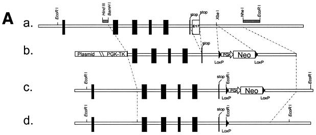
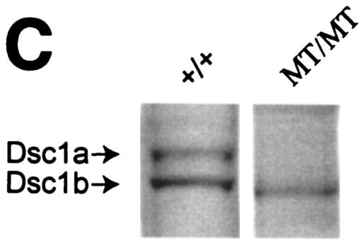
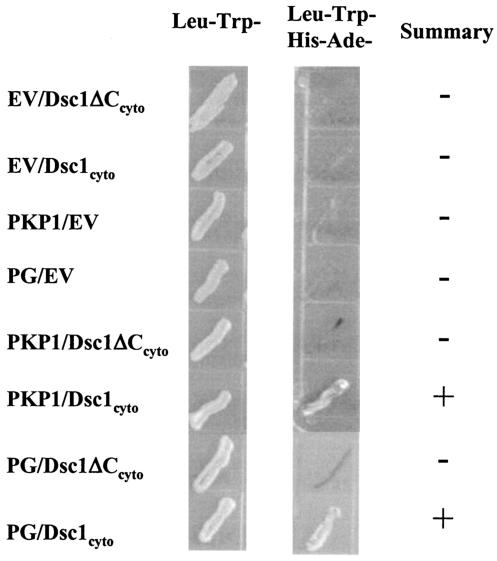
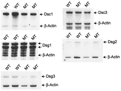
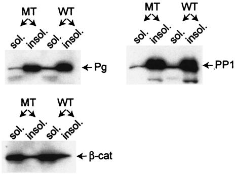
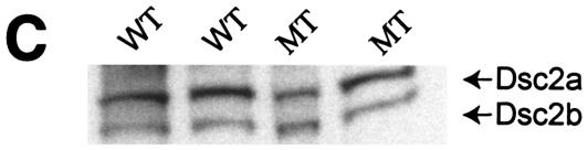
Desmocollin 1 (Dsc1) is part of a desmosomal cell adhesion receptor formed in terminally differentiating keratinocytes of stratified epithelia. The dsc1 gene encodes two proteins (Dsc1a and Dsc1b) that differ only with respect to their COOH-terminal cytoplasmic amino acid sequences. On the basis of in vitro experiments, it is thought that the Dsc1a variant is essential for assembly of the desmosomal plaque, a structure that connects desmosomes to the intermediate filament cytoskeleton of epithelial cells. We have generated mice that synthesize a truncated Dsc1 receptor that lacks both the Dsc1a- and Dsc1b-specific COOH-terminal domains. This mutant transmembrane receptor, which does not bind the common desmosomal plaque proteins plakoglobin and plakophilin 1, is integrated into functional desmosomes. Interestingly, our mutant mice did not show the epidermal fragility previously observed in dsc1-null mice. This suggests that neither the Dsc1a- nor the Dsc1b-specific COOH-terminal cytoplasmic domain is required for establishing and maintaining desmosomal adhesion. However, a comparison of our mutants with dsc1-null mice suggests that the Dsc1 extracellular domain is necessary to maintain structural integrity of the skin.


Figures

FIG. 1.
FIG. 1.
Strategy used to generate dsc1−/−ΔΕ17LoxP mutant mice. (A) Schematic representation of the targeting strategy used in this study. (a) The 3′ end of the mouse dsc1 gene locus is shown. Exons are represented by vertical bars. Exons 16 and 17 contain stop codons. Probes used to identify recombinant ES cell clones are shown as horizontal bars (HindIII/BamHI and NheI/EcoRI fragments). (b) Targeting construct TDSC1aΔE17. Intron 16 and exon 17 were deleted in this construct. A neomycin resistance minigene with flanking loxP sequences was inserted downstream of exon 17. The targeting vector also contained a thymidine kinase (TK) cassette. (c) Homologous recombination in ES cells generated the dsc1ΔE17Neo gene locus, in which intron 16 and exon 17 were deleted. (d) The neomycin resistance minigene was deleted through transient expression of CRE recombinase, generating the dsc1ΔE17LoxP allele. (B) RNase protection assays to detect Dsc1 RNA. Desmoglein 3 (left panel) and β-actin (right panel) were used as internal controls. Probes derived from dsc1 exon 17 (E17) and the 5′ end of the Dsc1 mRNA (5′), respectively, were used. Homozygous dsc1−/−ΔE17LoxP mutants express a Dsc1 RNA that does not contain exon 17 sequences. The genotype of the samples is indicated above the lanes (MT, dsc1−/−ΔE17LoxP mutant; +/+, wild type). Note that the expression levels in homozygous mutant mice are slightly lower than those in wild-type mice. (C) Western blot analysis using whole-skin extracts from wild-type (+/+) and homozygous mutant (MT/MT) mice with antibody gp899 (Dsc1). The positions of Dsc1a and Dsc1b in the wild-type sample are indicated. Note the absence of the Dsc1a band in the mutant sample.
FIG. 1.
FIG. 1.
Strategy used to generate dsc1−/−ΔΕ17LoxP mutant mice. (A) Schematic representation of the targeting strategy used in this study. (a) The 3′ end of the mouse dsc1 gene locus is shown. Exons are represented by vertical bars. Exons 16 and 17 contain stop codons. Probes used to identify recombinant ES cell clones are shown as horizontal bars (HindIII/BamHI and NheI/EcoRI fragments). (b) Targeting construct TDSC1aΔE17. Intron 16 and exon 17 were deleted in this construct. A neomycin resistance minigene with flanking loxP sequences was inserted downstream of exon 17. The targeting vector also contained a thymidine kinase (TK) cassette. (c) Homologous recombination in ES cells generated the dsc1ΔE17Neo gene locus, in which intron 16 and exon 17 were deleted. (d) The neomycin resistance minigene was deleted through transient expression of CRE recombinase, generating the dsc1ΔE17LoxP allele. (B) RNase protection assays to detect Dsc1 RNA. Desmoglein 3 (left panel) and β-actin (right panel) were used as internal controls. Probes derived from dsc1 exon 17 (E17) and the 5′ end of the Dsc1 mRNA (5′), respectively, were used. Homozygous dsc1−/−ΔE17LoxP mutants express a Dsc1 RNA that does not contain exon 17 sequences. The genotype of the samples is indicated above the lanes (MT, dsc1−/−ΔE17LoxP mutant; +/+, wild type). Note that the expression levels in homozygous mutant mice are slightly lower than those in wild-type mice. (C) Western blot analysis using whole-skin extracts from wild-type (+/+) and homozygous mutant (MT/MT) mice with antibody gp899 (Dsc1). The positions of Dsc1a and Dsc1b in the wild-type sample are indicated. Note the absence of the Dsc1a band in the mutant sample.
FIG. 2.
FIG. 2.
Characterization of dsc1 transcripts in newborn epidermis of wild-type (WT) and homozygous mutant (MT; dsc1−/−ΔE17LoxP) mice by RT-PCR. The 5′ primer was derived from the junction between exons 14 and 15, i.e., designed to suppress amplification of genomic DNA. The 3′ primer was derived from exon 16. (A) Agarose gel with RT-PCR products derived from three wild-type and three mutant samples. The PCR products were sequenced. The exon-intron organization of the product is schematically shown. (B) dsc1 gene structure and mRNA in wild-type and mutant mice. Note that exons 16 and 17 both contain stop codons. In mutant mice, we detected a single transcript that was aberrantly spliced, i.e., retained intron 15. DNA sequence analysis predicted that the mutant (MT) transcript encodes a protein in which the Dsc1b-specific sequence ESIRGHTLIKN is replaced by the sequence VSAQSSSAHSVQC. The sequence RLGE is encoded by the 3′ end of exon 15. Note that we have not confirmed the presence of the aberrant amino acid sequence at the COOH terminus of mutant Dsc1 by protein sequencing.
FIG. 3.
FIG. 3.
Molecular interactions of the cytoplasmic domains of Dsc1a (Dsc1cyto) and the truncated Dsc1 receptor (Dsc1ΔCcyto) synthesized in dsc1−/−ΔE17LoxP mutant mice. Yeast two-hybrid experiments were carried out to test for direct interactions by using growth in the absence of histidine and adenine (middle column) as a reporter for interactions between the cytoplasmic domains of the two Dsc1 molecules and several components of the desmosomal plaque. The cytoplasmic domain of Dsc1a, but not the COOH-terminally truncated Dsc1 mutant, interacts directly with the head domain of PKP1 and plakoglobin. Both Dsc polypeptides (Dsc1cyto and Dsc1ΔCcyto) were expressed as fusion proteins with the Gal4 activation domain and tested for interactions with either empty DNA binding domain vector (EV), the head domain of PKP1, or plakoglobin (PG) cloned into the Gal4 DNA binding domain vector.
FIG. 4.
FIG. 4.
Subcellular localization of Dsc1 in newborn epidermis of wild-type (WT) and mutant (MT) mice. (A) Deconvolution microscopy. The sections were stained with antibody gp899 (Dsc1; red) and a monoclonal antibody against desmoplakin (green). Colocalization resulted in yellow fluorescence. Note that the mutant protein coassembles with desmoplakin into desmosomes. Identical results were obtained by staining with gp899 and a monoclonal antibody against Dsg1 and Dsg2 (DG3.10; data not shown). (B) Low-temperature immunoelectron microscopy. Mutant (a, c, e, g) and wild-type (b, d, f, h) samples were incubated with various antibodies. (a, b) gp899 (silver enhancement; see Materials and Methods). (c, d) Higher magnification of desmosomes stained with gp899. (e, f) Costaining with gp899 (5-nm gold particles) and desmoplakin antibodies (15-nm gold particles). (g, h) Costaining with gp899 (5-nm gold particles) and plakoglobin antibodies (15-nm gold particles). Note that the mutant Dsc1 receptor is integrated into desmosomes and that the staining patterns of desmoplakin and plakoglobin are not affected by the mutation. Bars: a and b, 1 μm; c to h, 0.1 μm.
FIG. 5.
FIG. 5.
Western blot analysis of total-tissue lysates from back skin of newborn wild-type (WT) and dsc1−/−ΔE17LoxP mutant (MT) mice. Equal amounts of protein were blotted with antibodies against Dsg1 and Dsg2 (DG3.10), Dsg3, Dsc3 (gp2280; see Materials and Methods), plakoglobin (Pg), PKP1 (PP1), PKP3 (PP3), and β-catenin (β-cat). No significant differences in the expression levels of the analyzed proteins in wild-type and mutant samples were observed.
FIG. 6.
FIG. 6.
RNase protection assays to determine the expression levels of desmosomal cadherins in wild-type (WT) and dsc1−/−ΔE17LoxP mutant (MT) mice. RNA was isolated from the back epidermis of newborn mice. β-Actin served as an internal control in each experiment. Normalization of the expression data revealed that none of the markers showed a significant change (>50%) in their expression levels due to the dsc1 mutation. Note that the Dsg1 probe yielded two bands, which is probably due to a mouse strain polymorphism. The Dsg1 probe was derived from C57BL/6 genomic DNA. The mutant and wild-type samples tested were derived from mice on a segregating C57/BL6 and 129/SV background.
FIG. 7.
FIG. 7.
Western blot analysis of the TX-soluble (sol.) and -insoluble (insol.) protein fractions from the back skin of newborn wild-type (WT) and dsc1−/−ΔE17LoxP mutant (MT) mice. Note that plakoglobin (Pg) and PKP1 (PP1) are mainly found in the insoluble fraction, which is characteristic for junction-associated proteins. β-Catenin, on the other hand, is present predominantly in the soluble cytoplasmic pool. Again, no significant difference was observed between wild-type and mutant samples.
FIG. 8.
FIG. 8.
Effects of the dsc1 mutation on the expression of Dsc2. (A) RPA analysis of epidermal RNA from newborn wild-type (WT) and dsc1−/−ΔE17LoxP mutant (MT) samples. Note the dramatic increase in Dsc2 expression (approximately 38-fold) in the two mutant samples. β-Actin was used as an internal standard to normalize the expression data. (B) In situ hybridization with Dsc2 antisense probes. Note the strong suprabasal expression of Dsc2 in the mutant sample. The signal obtained with wild-type samples was barely above the background. A sense probe did not yield a signal on mutant or wild-type samples (data not shown). The dotted line indicates the position of the basement membrane. (C) Western blot analysis using total skin extracts from newborn mice with antibody gp2295. Note that Dsc2a and Dsc2b are expressed at similar levels in wild-type and homozygous mutant samples.
FIG. 8.
FIG. 8.
Effects of the dsc1 mutation on the expression of Dsc2. (A) RPA analysis of epidermal RNA from newborn wild-type (WT) and dsc1−/−ΔE17LoxP mutant (MT) samples. Note the dramatic increase in Dsc2 expression (approximately 38-fold) in the two mutant samples. β-Actin was used as an internal standard to normalize the expression data. (B) In situ hybridization with Dsc2 antisense probes. Note the strong suprabasal expression of Dsc2 in the mutant sample. The signal obtained with wild-type samples was barely above the background. A sense probe did not yield a signal on mutant or wild-type samples (data not shown). The dotted line indicates the position of the basement membrane. (C) Western blot analysis using total skin extracts from newborn mice with antibody gp2295. Note that Dsc2a and Dsc2b are expressed at similar levels in wild-type and homozygous mutant samples.

References

    1. Allen, E., Q. C. Yu, and E. Fuchs. 1996. Mice expressing a mutant desmosomal cadherin exhibit abnormalities in desmosomes, proliferation, and epidermal differentiation. J. Cell Biol. 133:1367-1382. - PMC - PubMed
    1. Amagai, M., N. Matsuyoshi, Z. H. Wang, C. Andl, and J. R. Stanley. 2000. Toxin in bullous impetigo and staphylococcal scalded-skin syndrome targets desmoglein 1. Nat. Med. 6:1275-1277. - PubMed
    1. Arnemann, J., K. H. Sullivan, A. I. Magee, I. A. King, and R. S. Buxton. 1993. Stratification-related expression of isoforms of the desmosomal cadherins in human epidermis. J. Cell Sci. 104(Pt. 3):741-750. - PubMed
    1. Barker, N., and H. Clevers. 2000. Catenins, Wnt signaling and cancer. Bioessays 22:961-965. - PubMed
    1. Bierkamp, C., K. J. McLaughlin, H. Schwarz, O. Huber, and R. Kemler. 1996. Embryonic heart and skin defects in mice lacking plakoglobin. Dev. Biol. 180:780-785. - PubMed

Publication types