. 2004 Jan;24(1):475-86.
doi: 10.1128/MCB.24.1.475-486.2004.

Dual effects of IkappaB kinase beta-mediated phosphorylation on p105 Fate: SCF(beta-TrCP)-dependent degradation and SCF(beta-TrCP)-independent processing

Affiliations

Dual effects of IkappaB kinase beta-mediated phosphorylation on p105 Fate: SCF(beta-TrCP)-dependent degradation and SCF(beta-TrCP)-independent processing

Shai Cohen et al. Mol Cell Biol. 2004 Jan.

Abstract

Processing of the p105 NF-kappaB precursor to yield the p50 active subunit is a unique and rare case in which the ubiquitin system is involved in limited processing rather than in complete destruction of its target. The mechanisms involved in this process are largely unknown, although a glycine repeat in the middle of p105 has been identified as a processing stop signal. IkappaB kinase (IKK)beta-mediated phosphorylation at the C-terminal domain with subsequent recruitment of the SCF(beta-TrCP) ubiquitin ligase leads to accelerated processing and degradation of the precursor, yet the roles that the kinase and ligase play in each of these two processes have not been elucidated. Here we demonstrate that IKKbeta has two distinct functions: (i) stimulation of degradation and (ii) stimulation of processing. IKKbeta-induced degradation is dependent on SCF(beta-TrCP), which acts through multiple lysine residues in the IkappaBgamma domain. In contrast, IKKbeta-induced processing of p105 is beta-transduction repeat-containing protein (beta-TrCP) independent, as it is not affected by expression of a dominant-negative beta-TrCP or following its silencing by small inhibitory RNA. Furthermore, removal of all 30 lysine residues from IkappaBgamma results in complete inhibition of IKK-dependent degradation but has no effect on IKK-dependent processing. Yet processing still requires the activity of the ubiquitin system, as it is inhibited by dominant-negative UbcH5a. We suggest that IKKbeta mediates its two distinct effects by affecting, directly and indirectly, two different E3s.


Figures

FIG. 1.
FIG. 1.
Schematic representation of p105-WT and the different p105 mutants. The different p105 proteins (a to i) were constructed and designated as described in Materials and Methods and in Table 1. Numbers in square brackets denote the amino acid residue number in the protein sequence. Numbers above the scheme line mark, in serially increasing numbers, all lysine residues that reside downstream of the GRR (downstream of lysine 425 [denoted 1]). The site of important restriction enzymes is marked (amino acid residue and enzyme name) in panels a and e. The different domains of p105 (RD, IKK phosphorylation and β-TrCP recognition sites, and the GRR) are marked and annotated in the figure body.
FIG. 2.
FIG. 2.
Truncated p105 proteins that span the IκBγ domain but lack the p50 portion of the molecule are efficiently ubiquitinated following phosphorylation. (A) In vitro-translated and [35S]methionine-labeled IKKβ-phosphorylated WT and p105-Δ917-933 were subjected to in vitro conjugation in a reconstituted cell-free system as described in Materials and Methods. SCFβ-TrCP was added where indicated. (B) [35S]methionine-labeled p105 mutants p105-577/END(577-968) and p105-660/END(660-986) were subjected to phosphorylation and ubiquitination as described in Materials and Methods and in the legend to panel A. (C) [35S]methionine-labeled p105-660/END(660-968) was preincubated in the absence or presence of IKKβ, and incubation continued in the presence of SCFβ-TrCP. Proteins were resolved by SDS-PAGE and were visualized by exposure to a PhosphorImager screen. Conj., conjugates.
FIG. 3.
FIG. 3.
Multiple p105 C-terminal lysine residues are targeted by β-TrCP. In vitro-translated, l-[35S]methionine-labeled, and IKKβ-phosphorylated WT and C-terminal K-to-R p105 mutants (see Table 1) were subjected to in vitro conjugation in a reconstituted cell-free system as described in Materials and Methods. Proteins were resolved by SDS-PAGE and were visualized by using a PhosphorImager. (A) Labeled p105-WT and the different K18-30R mutants (starting from p105-K18-23R; Table 1) were subjected to conjugation and were resolved by SDS-10% PAGE. (B) Data from three independent experiments, with the error bar representing one standard deviation, are presented. Percent conjugation is the ratio between radioactivity in the conjugates divided by the sum of the radioactivity in the conjugates and that in the remaining unreacted protein. (C) Reaction mixtures were similar to those described for panel A, except that ubiquitin was replaced with MeUb, and proteins were resolved via SDS-15% PAGE. Conj., conjugates.
FIG. 4.
FIG. 4.
Stimulation-induced processing and degradation of p105 is not affected by mutating lysine residues 18 (683) to 30 (967) in the C-terminal domain of the molecule. (A) COS-7 cells were transiently transfected with either a cDNA coding for p105-WT (lanes 2 to 5) or p105-K18-30R (lanes 6 to 9) as indicated. Control cells (lane 1) were transfected with an empty vector. Where indicated, a cDNA coding for constitutively active IKKβ was cotransfected. Twenty-four hours after transfection cells were harvested, and nuclear and cytosolic fractions were isolated as described in Materials and Methods. Aliquots of cytosolic and nuclear extracts representing an equal number of cells were resolved via SDS-10% PAGE and were blotted onto nitrocellulose paper, and proteins were visualized by using anti-p50 antibody and ECL as described in Materials and Methods. C, cytosolic fraction; N, nuclear fraction. (B) COS-7 cells were transiently transfected with a cDNA coding for either p105-WT (lanes 1 to 4), p105-K18-30R (lanes 5 to 8), or p105-Δ917-932 (lanes 9 to 12). Where indicated, cDNA coding for a constitutively active IKKβ was cotransfected. Twenty-four hours after transfection cells were pulse labeled with [35S]methionine (0; pulse). Following removal and dilution of the labeled amino acid and further incubation for 2 h (2; chase), the labeled proteins were immunoprecipitated by using anti-p50 antibody, resolved by SDS-10% PAGE, and visualized by phosphorimaging as described in Materials and Methods.
FIG. 5.
FIG. 5.
Removal of p105 lysine residues 12 to 30 reveals two distinct roles for IKK in p105 activation: stimulation of degradation and, independently, stimulation of processing. (A) Removal of lysine residues 1 (425) to 30 (969) completely inhibits conjugation of p105. In vitro-translated, [35S]methionine-labeled, and IKKβ-phosphorylated WT-p105 as well as p105 species that lack lysine residues 12 to 17 (p105-Δ544-654/ΔK12-17), 2 to 17 (p105-Δ429-654/ΔK2-17), 12 to 30 (p105-Δ544-654/ΔK12-17;K18-30R), and 1 to 30 (p105-Δ429-654/ΔK2-17;K1,18-30R) were subjected to SCFβ-TrCP-mediated conjugation in a cell-free system as described in Materials and Methods and in the legends to Fig. 2 and 3. Proteins were resolved by SDS-10% PAGE and were visualized by exposure to a PhosphorImager screen. Conj., conjugates. (B) Removal of p105 lysine residues 12 to 30 inhibits IKK-stimulated degradation but does not affect IKK-induced processing (Western blot analysis). COS-7 cells were transiently transfected with a cDNA coding for p105-Δ544-654/ΔK12-17 (lacking lysine residues 12 to 17; lanes 1 to 4) or p105-Δ544-654/ΔK12-17;K18-30R (lacking lysine residues 12 to 30; lanes 5 to 8). Where indicated, a cDNA coding for constitutive active IKKβ was cotransfected. Twenty-four hours after transfection cells were harvested and p105 and p50 were detected by Western blot analysis in nuclear and cytoplasmic fractions as described in Materials and Methods and in the legend to Fig. 4A. (C) Removal of p105 lysine residues 12 to 30 reduces IKK-stimulated degradation but does not affect induced processing (analysis by pulse-chase labeling and immunoprecipitation). COS-7 cells were transiently transfected for 24 h with cDNAs coding for the different species of p105 mutants and IKKβ as described in the legend to panel B. Processing and degradation of the p105 mutants was monitored in pulse-chase labeling and immunoprecipitation experiments as described in Materials and Methods and in the legend to Fig. 4B. Quantified data are presented. Percent of processing was calculated with the following equation: [(p50 detected at chase time − p50 detected at pulse time) × 1.74 (this value represents the net p50 generated during chase)/(p105 generated during pulse)] × 100. Percent of degradation was calculated with the following equation: {[p105 generated during pulse − p105 remaining after chase − (net p50 generated during chase × 1.74)]/p105 generated during pulse} × 100. Multiplication of the radioactivity in p50 by 1.74 was done in order to correct for the ratio of cysteine and methionine residues between p105 and p50. (D) Removal of p105 lysine residues 1 to 30 completely inhibits IKKβ-induced degradation, while processing remains unaffected. COS-7 cells were transiently transfected for 24 h with cDNAs coding for p105-WT (containing lysine residues 1 [425] to 30 [967]; lanes 1 to 4), p105-Δ429-654/ΔK2-17 (lacking lysine residues 2 to 17; lanes 5 to 8), or p105-Δ429-654/ΔK2-17;K1,18-30R (lacking lysine residues 1 to 30; lanes 9 to 12). Where indicated, cDNA coding for constitutive active IKKβ was cotransfected. Processing and degradation of the different p105 mutants was monitored in a pulse-chase labeling and immunoprecipitation experiment as described in Materials and Methods and in the legend to Fig. 4B. Quantified data are also presented (for quantification, see the legend to panel C).
FIG. 6.
FIG. 6.
β-TrCP is essential for stimulated degradation but not for stimulated processing of p105. (A) Suppression of β-TrCP inhibits degradation but not processing of p105. HEK 293 cells were transfected by either an empty vector (lane 1) or with cDNA coding for p105-WT (lanes 2 to 5). cDNAs coding for IKKβ (lanes 3 to 5) and ΔF-box β-TrCP (lane 4) or oligonucleotide for siRNA silencing of β-TrCP1 and -2 (lane 5) were transfected as described in Materials and Methods. Forty-eight hours after transfection with siRNA and 24 h after transfection with all other cDNAs, cells were harvested with RIPA buffer. Extracts representing an equal number of cells were resolved by SDS-10% PAGE and were blotted onto nitrocellulose paper, and proteins were visualized by using anti-p50 antibody and ECL as described in Materials and Methods. (B) Utilization of a dominant-negative ΔF-box β-TrCP or of p105 mutants that lack lysine residues 12 to 30 or 1 to 30 inhibits IKK-induced degradation but does not affect IKK-stimulated processing of p105. HEK 293 cells were transfected with either an empty vector (lane 1) or with cDNAs coding for p105-WT (lanes 2 to 4), p105-Δ544-654/ΔK12-17 (lanes 5 to 7), p105-Δ544-654/ΔK12-17;K18-30R (lanes 8 and 9), or p105-Δ429-654/ΔK2-17;K1,18-30R (lanes 10 and 11). Where indicated, cDNAs coding for ΔF-box β-TrCP and/or constitutively active IKKβ were cotransfected. Twenty-four hours after transfection cells were harvested with RIPA buffer, and disappearance of p105 and generation of p50 were monitored by using Western blot analysis as described in Materials and Methods and the legend to Fig. 4A. The anti-p50 antibody used here was different from the one that was used in the other experiments in that it did not recognize the 90-kDa nonspecific immune-reactive protein (marked once in panel A). This band masks the low-molecular-mass p105 deletion mutants.
FIG. 7.
FIG. 7.
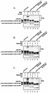
Signal-induced processing and degradation of p105 are dependent on an active ubiquitin-proteasome pathway. (A) Inhibition of the proteasome suppresses signal-induced processing and degradation of p105. COS-7 cells were transiently transfected for 24 h with a cDNA coding for p105-WT. Where indicated, cDNA coding for constitutive active IKKβ was cotransfected. Processing and degradation of p105 were monitored in a pulse-chase labeling and immunoprecipitation experiment in the absence (lanes 1 to 4) or presence (lanes 5 to 8) of lactacystin β-lactone, a 20S proteasome inhibitor, as described in Materials and Methods and in the legend to Fig. 4B. (B) The ubiquitin-conjugating enzymes UbcH5c and UbcH5a act differently on IKKβ-mediated processing of p105. COS-7 cells were transiently transfected with a cDNA coding for p105-WT. Where indicated, cDNAs coding for constitutively active IKKβ, DN-UbcH5a, or DN-UbcH5c were cotransfected. Twenty-four hours after transfection cells were harvested, and disappearance of p105 and generation of p50 were monitored by using Western blot analysis as described in Materials and Methods and in the legend to Fig. 4A. (C) The ubiquitin-conjugating enzymes UbcH5C and UbcH5A act differently on IKKβ-mediated processing of p105. This experiment is similar to the one described in Fig. 5B, except that it was carried out by using pulse-chase labeling and immunoprecipitation of p105 and p50 as described in Materials and Methods and in the legend to Fig. 4B. (D) Neither UbcH5c nor UbcH5a E2s are required for basal processing of p105. COS-7 cells were transiently transfected by either an empty vector (lane 1) or with cDNAs coding for p105-TthIII 1 (p105 to 544) (lanes 2 to 4). Where indicated, a cDNA coding for DN-UbcH5c (lane 3) or DN-UbcH5a (lane 4) was cotransfected. Twenty-four hours after transfection cells were harvested, and levels of p105 and p50 were monitored by using Western blot analysis as described in Materials and Methods and in the legend to Fig. 4A.
FIG. 8.
FIG. 8.
IKKβ-phosphorylated p105 mutant that lacks all 30 lysine residues in its IκBγ domain is ubiquitinated by a non-TrCP ubiquitin ligase. (A) In vitro-translated, [35S]methionine-labeled, and IKKβ-phosphorylated WT-p105 as well as p105 species that lack lysine residues 12 to 30 (p105-Δ544-654/ΔK12-17;K18-30R) and 1 to 30 (p105-Δ429-654/ΔK2-17;K1,18-30R) were subjected to SCFβ-TrCP-mediated conjugation in a cell-free system as described in Materials and Methods and in the legends to Fig. 2 and 3. (B) An experiment similar to the one described in panel A was carried out in crude HeLa extract rather then in a cell-free reconstituted system. Conjugation was monitored in the absence and presence of ATP. (C) An experiment the same as the one described in panel B was carried out, except that the [35S]methionine-labeled p105s were phosphorylated by IKK prior to their incubation in the HeLa extract. Proteins were resolved by SDS-10% PAGE and were exposed to a PhosphorImager screen as described in Materials and Methods and in the legends to Fig. 2 and 3. The lower panels show the different p105 species as observed under reduced exposure to demonstrate that equal amounts of radioactive proteins were loaded on the gels. Conj., conjugates.

References

    1. Alkalay, I., A. Yaron, A. Hatzubai, S. Jung, A. Avraham, O. Gerlitz, I. Pashut-Lavon, and Y. Ben-Neriah. 1995. In vivo stimulation of IκB phosphorylation is not sufficient to activate NF-κB. Mol. Cell. Biol. 15:1294-1301. - PMC - PubMed
    1. Baldi, L., K. Brown, G. Franzoso, and U. Siebenlist. 1996. Critical role for lysines 21 and 22 in signal-induced, ubiquitin-mediated proteolysis of IκBα. J. Biol. Chem. 271:376-379. - PubMed
    1. Bohuslav, J., V. V. Kravchenko, G. C. Parry, J. H. Erlich, S. Gerondakis, N. Mackman, and R. J. Ulevitch. 1998. Regulation of an essential innate immune response by the p50 subunit of NF-κB. J. Clin. Investig. 102:1645-1652. - PMC - PubMed
    1. Bradford, M. M. 1976. A rapid and sensitive method for the quantitation of microgram quantities of protein utilizing the principle of protein-dye binding. Anal. Biochem. 72:248-254. - PubMed
    1. Cohen, S., A. Orian, and A. Ciechanover. 2001. Processing of p105 is inhibited by docking of p50 active subunits to the ankyrin repeat domain, and inhibition is alleviated by signaling via the carboxyl-terminal phosphorylation/ubiquitin-ligase binding domain. J. Biol. Chem. 276:26769-26776. - PubMed

Publication types

MeSH terms