Comparative Study
. 2004 Feb 15;555(Pt 1):85-96.
doi: 10.1113/jphysiol.2003.056986. Epub 2003 Dec 12.

Cannabinoid-induced presynaptic inhibition at the primary afferent trigeminal synapse of juvenile rat brainstem slices

Affiliations
Comparative Study

Cannabinoid-induced presynaptic inhibition at the primary afferent trigeminal synapse of juvenile rat brainstem slices

Ying-Ching Liang et al. J Physiol. .

Abstract

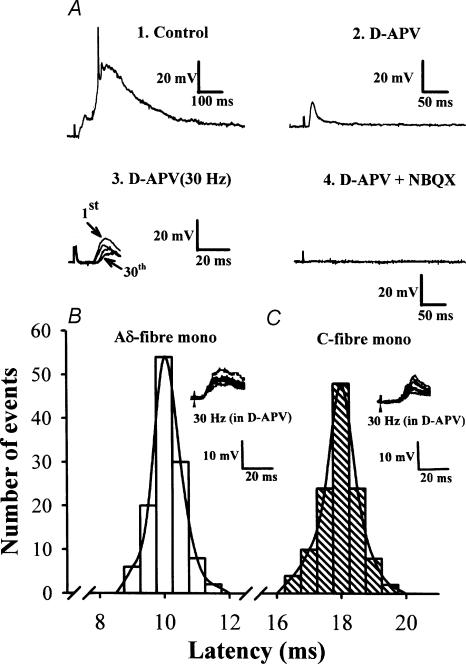
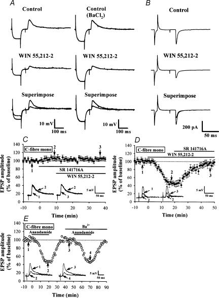
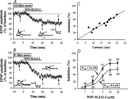
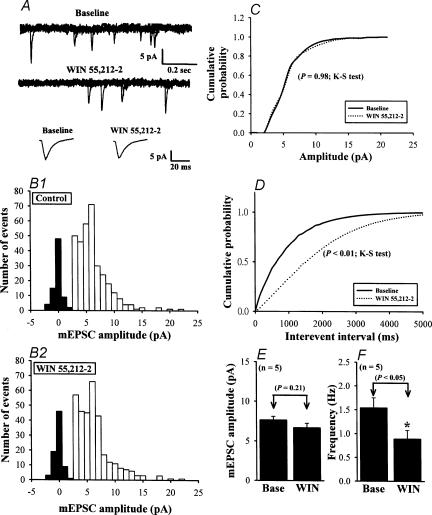
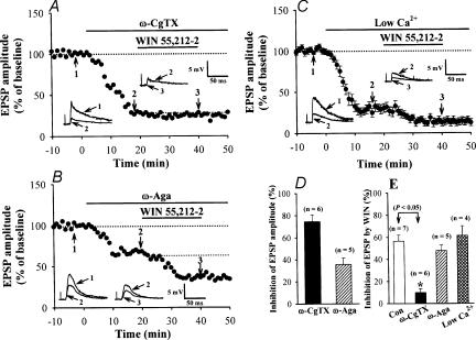
Systemic or intraventricular administration of cannabinoids causes analgesic effects, but relatively little is known for their cellular mechanism. Using brainstem slices with the mandibular nerve attached, we examined the effect of cannabinoids on glutamatergic transmission in superficial trigeminal caudal nucleus of juvenile rats. The exogenous cannabinoid receptor agonist WIN 55,212-2 (WIN), as well as the endogenous agonist anandamide, hyperpolarized trigeminal caudal neurones and depressed the amplitude of excitatory postsynaptic potentials (EPSPs) or currents (EPSCs) monosynaptically evoked by stimulating mandibular nerves in a concentration-dependent manner. The inhibitory action of WIN was blocked or fully reversed by the CB1 receptor antagonist SR 141716A. WIN had no effect on the amplitude of miniature excitatory postsynaptic currents (mEPSCs) recorded in the presence of tetrodotoxin or cadmium. The inhibitory effect of WIN on EPSCs was greater for those with longer synaptic latency, suggesting that cannabinoids have a stronger effect on C-fibre EPSPs than on Adelta-fibre EPSPs. Ba2+ (100 microm) blocked the hyperpolarizing effect of cannabinoids, but did not affect their inhibitory effect on EPSPs. The N-type Ca2+ channel blocker omega-conotoxin GVIA (omega-CgTX) occluded the WIN-mediated presynaptic inhibition, whereas the P/Q-type Ca2+ channel blocker omega-agatoxin TK (omega-Aga) had no effect. These results suggest that cannabinoids preferentially activate CB1 receptors at the nerve terminal of small-diameter primary afferent fibres. Upon activation, CB1 receptors may selectively inhibit presynaptic N-type Ca2+ channels and exocytotic release machinery, thereby attenuating the transmitter release at the trigeminal nociceptive synapses.


Figures

Figure 1
Figure 1. EPSPs recorded from trigeminal caudal neurones in response to mandibular nerve stimulation
A, EPSPs evoked in the trigeminal caudal neurones by stimulating the mandibular nerve at 0.033 Hz (1, 2, and 4) and at 30 Hz (3) in the presence of strychnine sulphate (0.5 μm) and bicuculline methiodide (10 μm). Perfusing the slices with NMDA receptor antagonist d-APV (50 μm) largely attenuated slow EPSP components (2) and further addition of the non-NMDA receptor antagonist NBQX (20 μm) completely abolished the synaptic response (4). Note that the short-latency fast EPSP component recorded in the presence of d-APV (5 traces superimposed) persisted up to 30 Hz with a relatively stable latency with no failures (3). The resting membrane potential of this cell was –67 mV. B and C, monosynaptic nature of EPSPs remaining after high-frequency repetitive stimulation in the presence of d-APV (50 μm). The latency histograms of EPSPs evoked at 30 Hz in two different trigeminal neurones are shown. From the stable latency and absence of failure during 30 Hz stimulation, we deduced that these afferent inputs are monosynaptically connected. Inset in B, representative monosynaptic Aδ-fibre EPSPs (6 traces superimposed) in one neurone had a low threshold (4 V; 0.3 ms) and short latency (10.6 ms). Inset in C, representative monosynaptic C-fibre EPSPs (6 traces superimposed) in one neurone had a high threshold (11 V; 0.3 ms) and long latency (18.6 ms). The latencies (120 responses each in B and C) were fitted with a Gaussian distribution.
Figure 2
Figure 2. Cannabinoid receptor agonists reduce monosynaptic EPSPs and EPSCs evoked by mandibular nerve stimulation
A, sample traces showing synaptically evoked EPSPs from cells recorded before and 20 min after application of WIN (5 μm) in the absence (left) or presence of Ba2+ (100 μm) (right). The EPSP was preceded by a transient hyperpolarizing current pulse (0.1 nA, 100 ms) passed through the recording microelectrode to measure input resistance (IR) of postsynaptic neurones. Note that bath application of WIN decreased the amplitude of EPSPs, which was accompanied by a substantial decrease in membrane IR. In the presence of Ba2+, WIN was still able to depress EPSP amplitude without affecting membrane IR. The average resting membrane potential (RMP) and IR of 12 neurones tested were −66.9 ± 2.7 mV and 105.4 ± 3.5 MΩ, respectively, before WIN application, and −68.9 ± 2.5 mV and 99.6 ± 3.2 MΩ after WIN application. B, sample traces showing the effect of WIN (5 μm) on monosynaptic EPSCs. The neurone was voltage clamped at a holding potential of −70 mV, and EPSCs were evoked at 30 s intervals in the presence of strychnine sulphate (0.5 μm), bicuculline methiodide (10 μm) and d-APV (50 μm). Traces are averaged from 6 responses. C, summary time plot of C-fibre EPSPs (n= 5) showing a potentiating effect of SR 141716A (5 μm) on the EPSP amplitude and its blocking effect on the WIN-induced inhibition of EPSPs. D, summary time plot of C-fibre EPSPs (n= 4) showing that SR 141716A (5 μm) reversed the WIN-induced inhibition of EPSPs. E, effect of external Ba2+ on the anandamide-induced inhibition of EPSPs. Ba2+ (100 μm) had no effect on the inhibitory effect of anandamide (30 μm) on monosynaptic C-fibre EPSPs. Superimposed EPSPs were taken at the times indicated. Similar results were also observed in other three cells. Bars indicate the period of drug application.
Figure 3
Figure 3. Stronger inhibition by WIN of C-fibre EPSPs than Aδ-fibre EPSPs
A, averaged time course of the inhibitory effect of WIN (5 μm) on monosynaptic Aδ-fibre EPSPs (n= 5). B, averaged time course showing the effect of WIN on monosynaptic C-fibre EPSPs (n= 7). Insets show superimposed EPSPs taken at the indicated times (1–3). C, comparison between the magnitude of the WIN-induced EPSP inhibition (ordinate, 20 min after WIN application) and the synaptic latency of EPSPs (abscissa). A regression line was drawn using the least squares method (r= 0.89). D, dose–response curves for depression of Aδ- (◯) and C-fibre EPSPs (•) by WIN. The numbers in parentheses indicate the number of slices tested.
Figure 4
Figure 4. Effect of WIN on the mEPSCs
A, sample traces of mEPSCs before (Baseline: in the presence of 1 μm TTX to block Na+ channels) and during application of 5 μm WIN. Lower traces are averaged mEPSCs of 35 events each before and after WIN application, demonstrating the lack of effect of WIN on the amplitude and kinetics of mEPSCs. B, amplitude histograms of mEPSCs before (B1) and during WIN application (B2) obtained from 323 and 312 mEPSC events, respectively. C, cumulative probability plots of the mEPSC amplitude before (dotted line) and during (continuous line) application of WIN. No changes occurred in the distribution during WIN application (P= 0.98; Kolmogorov-Smirnov test). D, cumulative interevent interval distribution, illustrating a significant increase in the interevent interval (i.e. decreased frequency; P < 0.01; Kolmogorov-Smirnov test) during WIN application. Data in AD were obtained from the same neurone. E and F, summary of the effect of 5 μm WIN on the average amplitude (E) and frequency (F) of mEPSCs (n= 5). Data are presented as means ±s.e.m. Asterisk indicates P < 0.05 compared with control (Base). Holding potential was −70 mV.
Figure 5
Figure 5. Involvement of presynaptic N-type VACCs in WIN-induced synaptic inhibition
A, a typical experiment in which the slice was perfused with 1 μmω-CgTX, which blocked ∼74% of the monosynaptic C-fibre EPSPs. In the presence of ω-CgTX, WIN (5 μm) no longer attenuated EPSPs. B, a typical experiment in which the slice was perfused with 200 nmω-Aga-TK, which blocked ∼34% of the monosynaptic C-fibre EPSPs. The fraction of EPSPs insensitive to ω-Aga was still sensitive to WIN. C, average time course (n= 4) showing the WIN-induced depression of the monosynaptic C-fibre EPSPs in a low Ca2+ solution. The standard ACSF was replaced by a low-Ca2+ solution containing 0.8 mm Ca2+ and 2.9 mm Mg2+. The superimposed EPSPs in the insets in each graph illustrate respective recordings from example experiments taken at the times indicated by the numbers (1–3). Horizontal bars indicate the period of application of drugs and low-Ca2+ solution. D, the magnitude of EPSP attenuation by Ca2+ channel blockers. E, summary of WIN (5 μm)-induced synaptic inhibition, in the presence of Ca2+ channel blockers or in a low-Ca2+ solution. Data are presented as means ±s.e.m. Asterisk indicates P < 0.05 in Student's unpaired t test.

References

    1. Ameri A. The effects of cannabinoids on the brain. Prog Neurobiol. 1999;58:315–348. - PubMed
    1. Ataka T, Kumamoto E, Shimoji K, Yoshimura M. Baclofen inhibits more effectively C-afferent than Aδ-afferent glutamatergic transmission in substantia gelatinosa neurons of adult rat spinal cord slices. Pain. 2000;86:273–282. - PubMed
    1. Auclair N, Otani S, Soubrie P, Crépel F. Cannabinoids modulate synaptic strength and plasticity at glutamatergic synapses of rat prefrontal cortex pyramidal neurons. J Neurophysiol. 2000;83:3287–3293. - PubMed
    1. Bao JP, Li JJ, Perl ER. Differences in Ca2+ channels governing generation of miniature and evoked excitatory synaptic currents in spinal laminae I and II. J Neurosci. 1998;18:8740–8750. - PMC - PubMed
    1. Basbaum AI, Jessell TM. The perception of pain. In: Kandel ER, Schwartz JH, Jessell TM, editors. Principles of Neural Science. 4. Columbus: McGraw-Hill Companies; 2000. pp. 472–491.

Publication types

MeSH terms