Review
. 2004 Mar;13(2):172-80.
doi: 10.1007/s00586-003-0653-5. Epub 2003 Dec 13.

Atlanto-occipital dislocation: four case reports of survival in adults and review of the literature

Affiliations
Review

Atlanto-occipital dislocation: four case reports of survival in adults and review of the literature

Ludwig Labler et al. Eur Spine J. 2004 Mar.

Abstract

Traumatic atlanto-occipital dislocation (AOD) is a rare cervical spine injury and in most cases fatal. Consequently, relatively few case reports of adult patients surviving this injury appeared in the literature. We retrospectively report four patients who survived AOD injury and were treated at our institution. A young man fell from height and a woman was injured in a traffic accident. Both patients survived the injury but died later in the hospital. The third patient had a motorcycle accident and survived with incomplete paraplegia. The last patient, a man involved in a working accident, survived without neurological deficit of the upper extremities. Rigid posterior fixation and complete reduction of the dislocation were applied in last two cases using Cervifix together with a cancellous bone grafting. Previously reported cases of patients surviving AOD are reviewed, and clinical features and operative stabilisation procedures are discussed.


Figures

Fig. 1
Fig. 1
Sagittal and frontal T2-weighted MRI sequences. Lesion in the pontomedullary junction (arrows)
Fig. 2
Fig. 2
Functional examination under image intensifier demonstrating atlanto-occipital dislocation (AOD)
Fig. 3 a
Fig. 3 a
Positioning of the patient, in prone position, with fixation of the head by a Mayfield clamp. b Postoperative X-rays showing stabilization of the AOD by dorsal fusion, from the occiput to C5, with Cervifix and cancellous bone
Fig. 4 a
Fig. 4 a
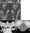
Atlanto-occipital dissociation in the CT scan demonstrating the subluxation with a rotationary component after first reduction (open arrow) and avulsion fracture of the ligaments (solid arrows). b A 3D reconstruction with an anteroposterior view (arrows). c Subluxation viewed cranially through the foramen magnum (arrows)
Fig. 5
Fig. 5
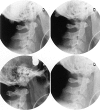
Functional tests of the atlanto-occipital junction under image intensifier
Fig. 6 a
Fig. 6 a
An MRI scan of AOD shows an epidural haematoma (arrows) with b associated lesion of the left vertebral artery (arrow)
Fig. 7
Fig. 7
Dorsal fusion from the occiput to C5 by Cervifix

References

    1. Adams J Forensic Sci. 1992;37:556. - PubMed
    1. Ahuja Surg Neurol. 1994;41:112. - PubMed
    1. Alker Orthop Clin North Am. 1978;9:1003. - PubMed
    1. Angel N Engl J Med. 2001;345:1256. doi: 10.1056/NEJMicm070559. - DOI - PubMed
    1. Babbitz Spine. 2001;26:1401. doi: 10.1097/00007632-200106150-00030. - DOI - PubMed