The extracellular matrix of porcine mature oocytes: origin, composition and presumptive roles
- PMID: 14675483
- PMCID: PMC317375
- DOI: 10.1186/1477-7827-1-124
The extracellular matrix of porcine mature oocytes: origin, composition and presumptive roles
Abstract
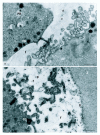
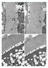
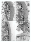
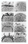
The extracellular matrix (ECM) of porcine mature oocytes was revealed by transmission electron microscopy (TEM) after treatment with tannic acid and ruthenium red. Present in the perivitelline space (PVS) and on the surface of the zona pellucida (ZP), it appeared to be composed of thin filaments and granules at the interconnections of the filaments, which were interpreted respectively as hyaluronic acid chains and bound proteoglycans. In order to determine whether this material is produced by the corona cells (the same ECM was found also on the surface of the zona pellucida and between cumulus cells) or by the oocyte itself, the synthesis of glycoproteins and glycosaminoglycans was checked by autoradiography on semi-thin and thin sections observed by light and electron microscopy. Immature oocytes within or without cumulus cells, were incubated with L [3H-] fucose or L [3H-] glucosamine--precursors respectively of glycoproteins and hyaluronic acid or hyaluronan (HA) bound to proteoglycans--for various times (with or without chase) and at different stages during in vitro maturation. In the first case, incorporation was found in both cumulus cells and ooplasm (notably in the Golgi area for 3H-fucose) and labeled material accumulated in the ECM of the PVS and of the ZP surface. Labeling in the PVS with both precursors was maximum between metaphase I (MI) and metaphase II (MII) and was partially extracted by hyaluronidase but not by neuraminidase. Tunicamycin, an inhibitor of glycoprotein synthesis, significantly decreased the amount of 3H-fucose labeled molecules in the PVS and increased the incidence of polyspermic penetration during subsequent in vivo fertilization. Since cumulus-free oocytes also secreted 3H-glucosamine containing compounds, both oocyte and cumulus cells probably contribute to the production of the ECM found in the PVS of mature oocytes. ECM and particularly its HA moiety present on both sides of the ZP may constitute a favourable factor for sperm penetration.
Figures


Similar articles
-
Structure, distribution and composition of the extracellular matrix of human oocytes and cumulus masses.Hum Reprod. 1992 Mar;7(3):391-8. doi: 10.1093/oxfordjournals.humrep.a137656. Hum Reprod. 1992. PMID: 1375234
-
Involvement of N-glycosylation of zona glycoproteins during meiotic maturation in sperm-zona pellucida interactions of porcine denuded oocytes.Anim Sci J. 2013 Jan;84(1):8-14. doi: 10.1111/j.1740-0929.2012.01027.x. Epub 2012 Jun 6. Anim Sci J. 2013. PMID: 23302076
-
N-glycosylation of zona glycoproteins during meiotic maturation is involved in sperm-zona pellucida interactions of porcine oocytes.Theriogenology. 2011 Apr 1;75(6):1146-52. doi: 10.1016/j.theriogenology.2010.11.026. Epub 2011 Jan 8. Theriogenology. 2011. PMID: 21220169
-
Zona pellucida characteristics and sperm-binding patterns of in vivo and in vitro produced porcine oocytes inseminated with differently prepared spermatozoa.Theriogenology. 2005 Jan 15;63(2):352-62. doi: 10.1016/j.theriogenology.2004.09.044. Theriogenology. 2005. PMID: 15626404 Review.
-
Regulation of cumulus expansion and hyaluronan synthesis in porcine oocyte-cumulus complexes during in vitro maturation.Endocr Regul. 2012 Oct;46(4):225-35. doi: 10.4149/endo_2012_04_225. Endocr Regul. 2012. PMID: 23127506 Review.
Cited by
-
Effect of D-Glucuronic Acid and N-acetyl-D-Glucosamine Treatment during In Vitro Maturation on Embryonic Development after Parthenogenesis and Somatic Cell Nuclear Transfer in Pigs.Animals (Basel). 2021 Apr 6;11(4):1034. doi: 10.3390/ani11041034. Animals (Basel). 2021. PMID: 33917537 Free PMC article.
-
Follicular extracellular vesicles enhance meiotic resumption of domestic cat vitrified oocytes.Sci Rep. 2020 May 25;10(1):8619. doi: 10.1038/s41598-020-65497-w. Sci Rep. 2020. PMID: 32451384 Free PMC article.
-
Sperm proteasomes degrade sperm receptor on the egg zona pellucida during mammalian fertilization.PLoS One. 2011 Feb 23;6(2):e17256. doi: 10.1371/journal.pone.0017256. PLoS One. 2011. PMID: 21383844 Free PMC article.
References
-
- Talbot P, Dicarlantonio G. The oocyte-cumulus complex: ultrastructure of the extracellular components in hamster and mice. Gamete Res. 1984;10:127–142.
-
- Talbot P. Sperm penetration through oocyte investments in mammals. Am J Anat. 1985;174:331–346. - PubMed
-
- Dandekar P, Aggeler J, Talbot P. Structure, distribution and composition of the extracellular matrix of human oocytes and cumulus masses. Hum Reprod. 1992;7:391–398. - PubMed
-
- Fléchon J-E. Application of cytochemical techniques to the study of maturation of gametes and fertilization in mammals. Multipurpose use of glycolmethacrylate embedding. Histochem J. 1974;6:65–67.
-
- Talbot P, Dicarlantonio G. Ultrastructure of opossum oocyte investing coats and their sensitivity to trypsin and hyaluronidase. Dev Biol. 1984;103:159–167. - PubMed
Publication types
MeSH terms
Substances
LinkOut - more resources
Full Text Sources
Research Materials

