

A role for arrays in clinical virology: fact or fiction?
- PMID: 14675863
- PMCID: PMC7128301
- DOI: 10.1016/j.jcv.2003.08.002
A role for arrays in clinical virology: fact or fiction?
Abstract
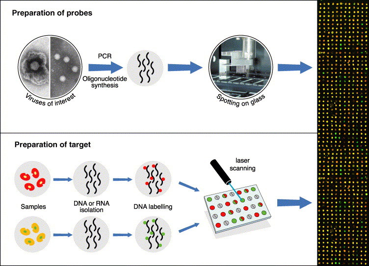
Microarrays of DNA probes have at least three roles in clinical virology. These are: firstly, in diagnosis, to recognise the causative agent of an illness; secondly, for molecular typing for (i) patient management, (ii) epidemiological reasons (e.g. investigating routes of transmission), (iii) purposes related to vaccine use; and thirdly, in research, to investigate the interactions between the virus and the host cell. Microarrays intended for syndromic diagnostic purposes require genome specific probes to capture the unknown target viral sequences and thereby reveal the presence of that virus in a test sample. Microarrays intended for typing and patient management, e.g. monitoring antiviral drug resistant mutations require a set of probes representing the important sequence variants of one or more viral genes. Microarrays intended for research into virus-host interactions require probes representative of each individual gene or mRNA of either the virus or the host genome. Diagnostic microarrays are dependent for their utility and versatility on generic, multiplex or random polymerase chain reactions that will amplify any of several (unknown) viral target sequences from a patient sample. In this review, the existing and potential applications of microarrays in virology, and the problems that need to be overcome for future success, are discussed.
Figures
Comment in
-
Regional virus laboratory Kelvin buildings.J Clin Virol. 2004 Jan;29(1):1. doi: 10.1016/j.jcv.2003.11.008. J Clin Virol. 2004. PMID: 14675862 No abstract available.
Similar articles
-
Molecular and diagnostic clinical virology in real time.Clin Microbiol Infect. 2004 May;10(5):471; author reply 471-2. doi: 10.1111/j.1469-0691.2004.00905.x. Clin Microbiol Infect. 2004. PMID: 15113330 Free PMC article. No abstract available.
-
Consolidation of molecular testing in clinical virology.Expert Rev Anti Infect Ther. 2017 Apr;15(4):387-400. doi: 10.1080/14787210.2017.1271711. Epub 2016 Dec 24. Expert Rev Anti Infect Ther. 2017. PMID: 28002969 Review.
-
Virome Capture Sequencing Enables Sensitive Viral Diagnosis and Comprehensive Virome Analysis.mBio. 2015 Sep 22;6(5):e01491-15. doi: 10.1128/mBio.01491-15. mBio. 2015. PMID: 26396248 Free PMC article.
-
Regional virus laboratory Kelvin buildings.J Clin Virol. 2004 Jan;29(1):1. doi: 10.1016/j.jcv.2003.11.008. J Clin Virol. 2004. PMID: 14675862 No abstract available.
-
Multiplex PCR: optimization and application in diagnostic virology.Clin Microbiol Rev. 2000 Oct;13(4):559-70. doi: 10.1128/CMR.13.4.559. Clin Microbiol Rev. 2000. PMID: 11023957 Free PMC article. Review.
Cited by
-
Diagnostic approaches for viruses and prions in stem cell banks.Virology. 2006 Mar 30;347(1):1-10. doi: 10.1016/j.virol.2005.11.026. Epub 2005 Dec 27. Virology. 2006. PMID: 16380145 Free PMC article. Review.
-
Virus discovery by sequence-independent genome amplification.Rev Med Virol. 2006 Nov-Dec;16(6):365-83. doi: 10.1002/rmv.515. Rev Med Virol. 2006. PMID: 16929467 Free PMC article. Review.
-
The Future of Gene Expression Studies in HIV Research.Curr HIV Res. 2025;23(1):28-34. doi: 10.2174/011570162X361179250204061607. Curr HIV Res. 2025. PMID: 39936446 Review.
-
Comparison of the digene HPV genotyping LQ test and the PANArray HPV genotyping chip for detection of high-risk or probable high-risk human papillomavirus genotypes.Ann Lab Med. 2014 Jul;34(4):279-85. doi: 10.3343/alm.2014.34.4.279. Epub 2014 Jun 19. Ann Lab Med. 2014. PMID: 24982832 Free PMC article.
-
Advances in viral respiratory infections: new experimental models.Drug Discov Today Dis Models. 2004 Dec 17;1(3):303-309. doi: 10.1016/j.ddmod.2004.11.011. Epub 2004 Dec 8. Drug Discov Today Dis Models. 2004. PMID: 32288769 Free PMC article. Review.
References
-
- Aberle S.W., Kletzmayr J., Watschinger B., Schmied B., Vetter N., Puchhammer-Stockl E. Comparison of sequence analysis and the INNO-LiPA HBV DR line probe assay for detection of lamivudine-resistant hepatitis B virus strains in patients under various clinical conditions. J. Clin. Microbiol. 2001;39:1972–1974. - PMC - PubMed
-
- Amexis G., Rubin S., Chizhikov V., Pelloquin F., Carbone K., Chumakov K. Sequence diversity of Jeryl Lynn strain of mumps virus: quantitative mutant analysis for vaccine quality control. Virology. 2002;300:171–179. - PubMed
-
- Bohlander S.K., Espinosa R., 3rd, Le Beau M.M., Rowley J.D., Diaz M.O. A method for the rapid sequence-independent amplification of microdissected chromosomal material. Genomics. 1992;13:1322–1324. - PubMed
-
- Boonham N., Walsh K., Smith P., Madagan K., Graham I., Barker I. Detection of potato viruses using microarray technology: towards a generic method for plant viral disease diagnosis. J. Virol. Meth. 2003;108:181–187. - PubMed
-
- Bugawan T.L., Apple R., Erlich H.A. A method for typing polymorphism at the HLA-A locus using PCR amplification and immobilized oligonucleotide probes. Tissue Antigens. 1994;44:137–147. - PubMed
Publication types
MeSH terms
Substances
LinkOut - more resources
Full Text Sources
Other Literature Sources
Medical
