. 2003 Dec 15;198(12):1887-97.
doi: 10.1084/jem.20030621.

The Helicobacter pylori vacuolating toxin inhibits T cell activation by two independent mechanisms

Affiliations

The Helicobacter pylori vacuolating toxin inhibits T cell activation by two independent mechanisms

Marianna Boncristiano et al. J Exp Med. .

Abstract

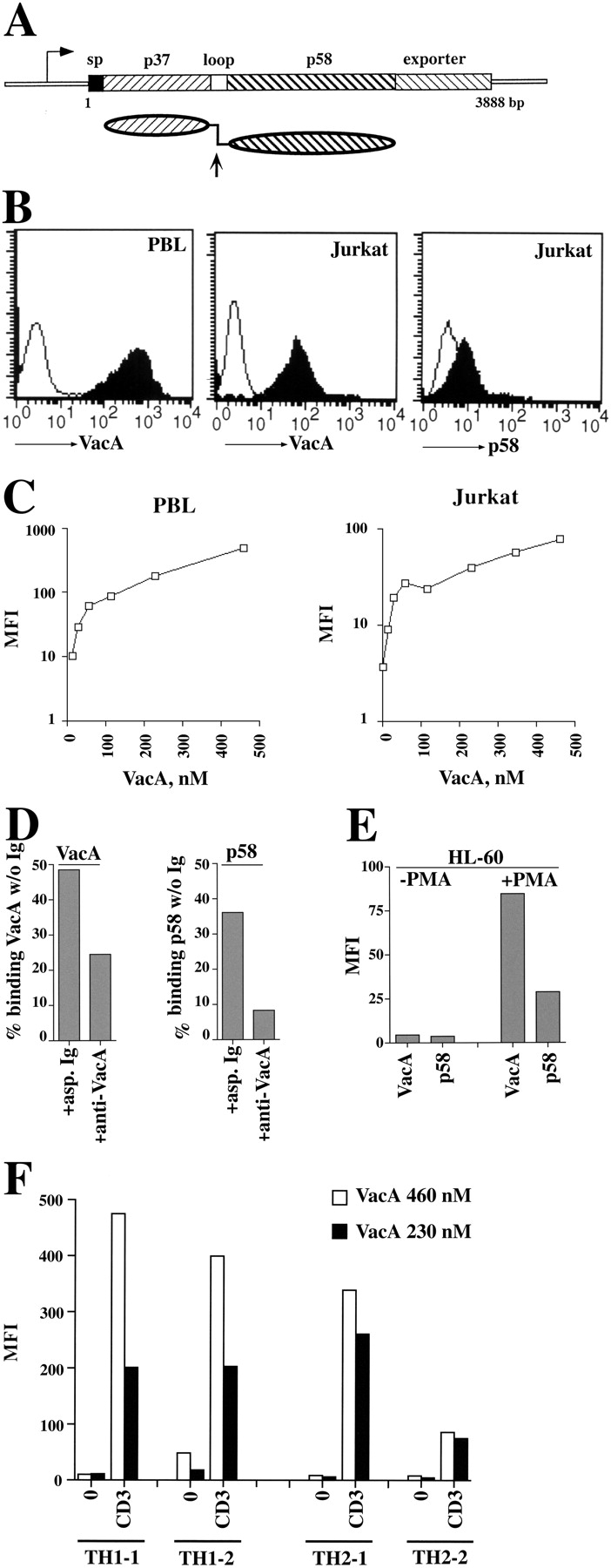
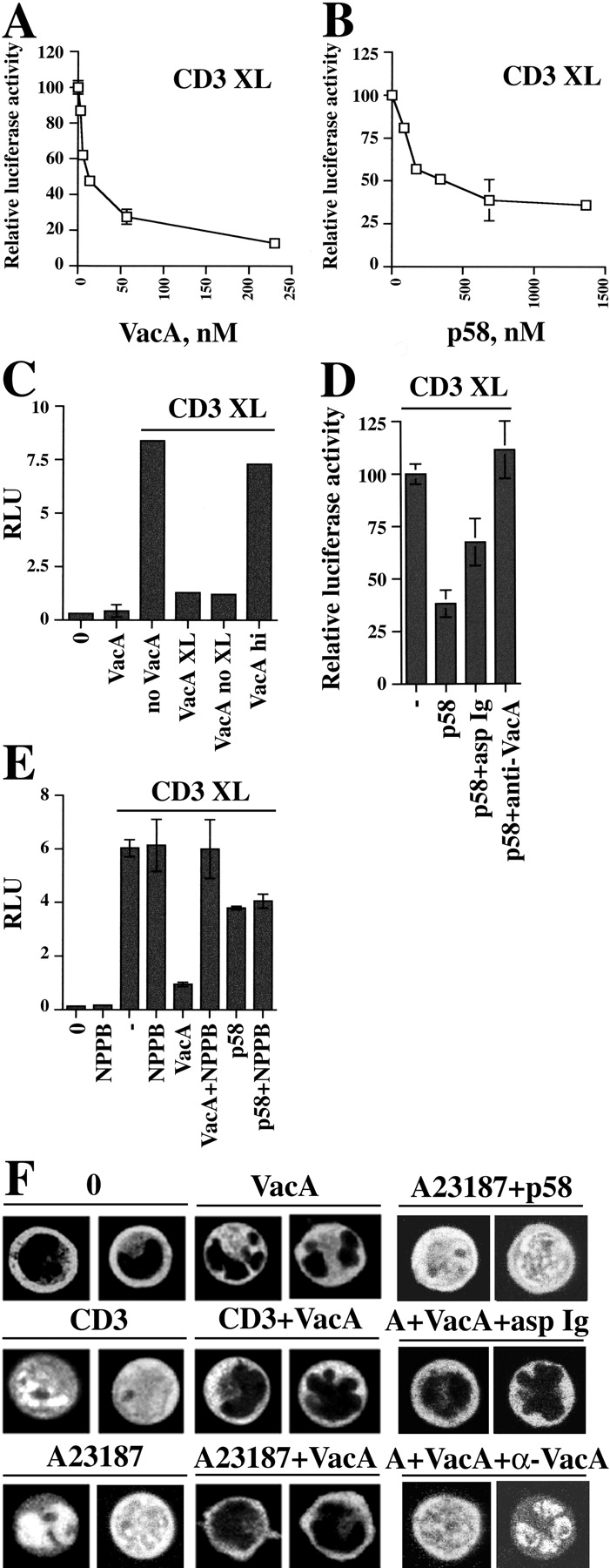
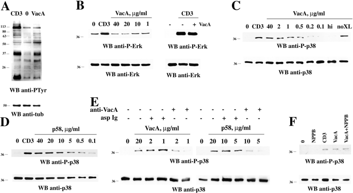
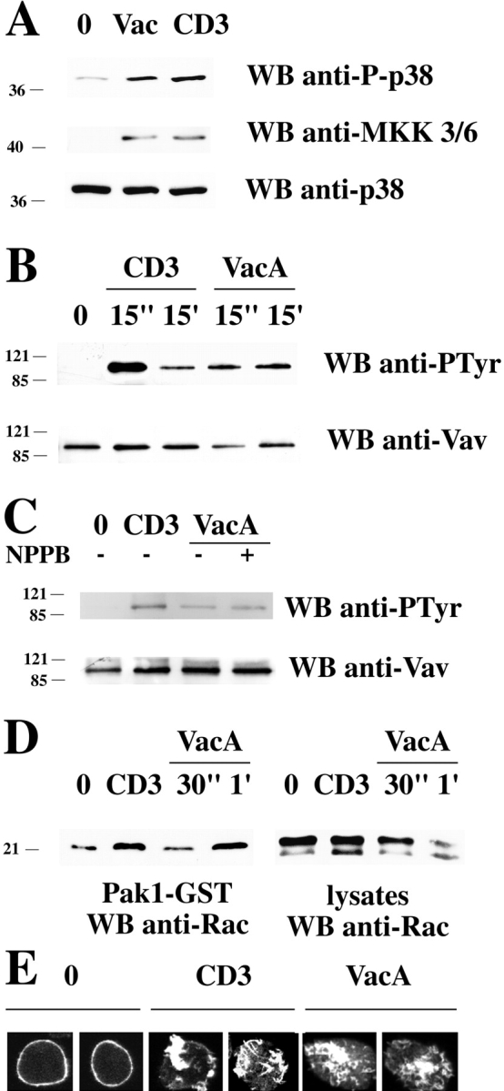
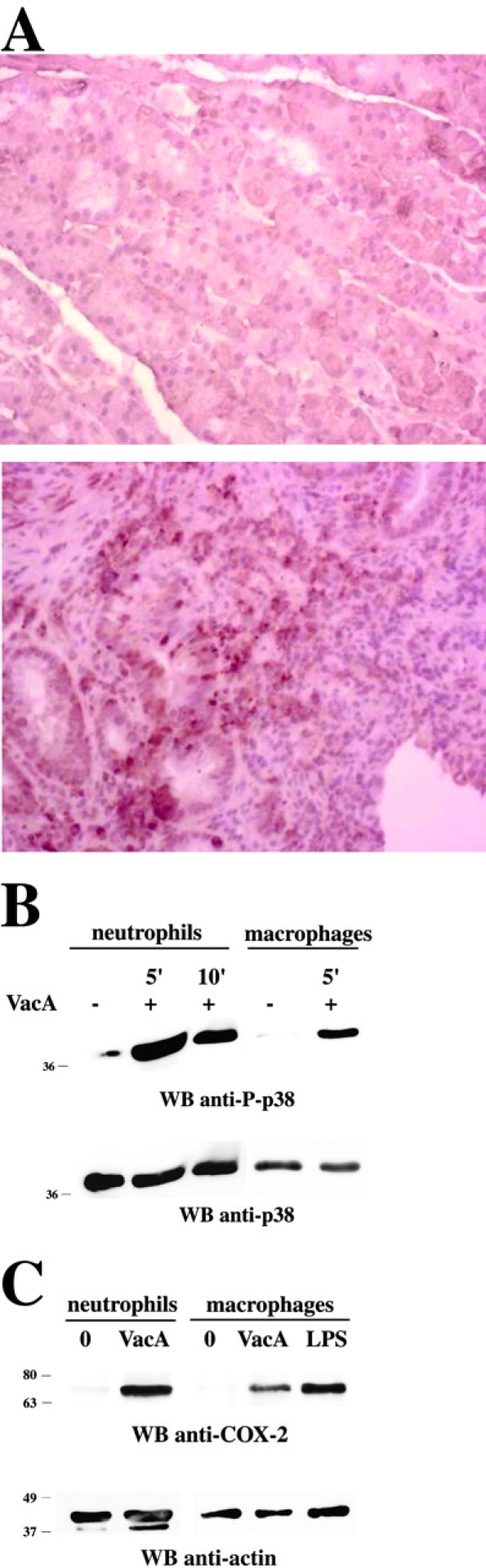
Helicobacter pylori toxin, VacA, damages the gastric epithelium by erosion and loosening of tight junctions. Here we report that VacA also interferes with T cell activation by two different mechanisms. Formation of anion-specific channels by VacA prevents calcium influx from the extracellular milieu. The transcription factor NF-AT thus fails to translocate to the nucleus and activate key cytokine genes. A second, channel-independent mechanism involves activation of intracellular signaling through the mitogen-activated protein kinases MKK3/6 and p38 and the Rac-specific nucleotide exchange factor, Vav. As a consequence of aberrant Rac activation, disordered actin polymerization is stimulated. The resulting defects in T cell activation may help H. pylori to prevent an effective immune response leading to chronic colonization of its gastric niche.


Figures

Figure 1.
Figure 1.
VacA binding on Jurkat T cells and human PBL and Th cells. (A) Schematic representation of the vacA gene and VacA protein. The 3,888-bp vacA gene from H. pylori strain CCUG17874 is represented showing regions encoding the signal peptide (sp), the 37-kD (p37) and 58-kD (p58) natural proteolytic fragments separated by the short hydrophilic region containing the cleavage site (loop), and the outer membrane autotransporter (exporter). Below is represented the mature secreted protein showing the two domains connected by a flexible loop. The arrow indicates the site of proteolytic cleavage between p37 and p58. (B) Flow cytometric analysis of VacA and p58 binding to purified human PBL (left) or Jurkat T cells (middle, right). VacA and p58 concentrations were 40 μg/ml (460 nM) and 80 μg/ml (1,365 nM), respectively. (C) Titration of VacA binding to PBL (left) or Jurkat cells (right) by flow cytometry. The data are expressed as mean fluorescence intensity (MFI). (D) Residual VacA or p58 binding to Jurkat cells after immunodepletion with either neutralizing anti-VacA or irrelevant (asp) Ig (starting VacA/p58 concentration 20 μg/ml, Ig 100 μg/ml). The data, obtained by flow cytometry, are expressed as the percentage of binding of the same concentration of untreated VacA/p58. (E) Flow cytometric analysis of VacA/p58 (40 μg/ml) binding to HL-60 cells, either untreated or incubated for 48 h with carrier or 20 ng/ml PMA. (F) Flow cytometric analysis of VacA binding to Th1 and Th2 cells, either unstimulated (0) or after 16-h stimulation with plastic-bound anti-CD3 mAb (CD3). Two representative Th1/Th2 clones are shown.
Figure 2.
Figure 2.
VacA inhibits the induction of CD69 surface expression in response to TCR–CD3 engagement. (A) Flow cytometric analysis of CD69 expression on Jurkat cells activated for 16 h by CD3 cross-linking in the presence or absence of either 20 μg/ml VacA or 40 μg/ml p58. (B) CD69 expression on human PBL activated for 24 h by CD3 cross-linking in the presence or the absence of 20 μg/ml VacA. Cells were counterstained with fluorochrome-labeled anti-CD4 or anti-CD8 mAb, and CD69 expression was measured on gated CD4+ (left) or CD8+ (right) T cells.
Figure 3.
Figure 3.
Inhibition of CD3-dependent NF-AT activation and nuclear translocation by VacA. Relative luciferase activity in NFAT/luciferase reporter Jurkat cells activated by CD3 cross-linking (CD3 XL) in the presence of different concentrations of VacA (A) or p58 (B). Luciferase activity induced by CD3 cross-linking in the absence of VacA was taken as 100%. (C) Relative luciferase units (RLU) in reporter Jurkat cells activated by CD3 cross-linking in the presence or absence of 20 μg/ml VacA. VacA was either cross-linked using anti-VacA mAb and plate-bound secondary antibodies (VacA XL) or bound to Jurkat cells and plated on secondary antibodies without anti-VacA mAb (VacA noXL). VacA hi, heat-inactivated VacA (boiled 5 min), cross-linked. (D) Relative luciferase activity (percentage of activity triggered by CD3 cross-linking) in reporter Jurkat cells activated by CD3 cross-linking in the presence or absence of 20 μg/ml p58. p58 was used either as such or after preincubation with 100 μg/ml neutralizing anti-VacA (p58+α-VacA) or irrelevant Ig (p58+asp Ig) and cross-linked using anti-VacA mAb and plate-bound secondary antibodies. (E) Relative luciferase units (RLU) in reporter Jurkat cells activated by CD3 cross-linking in the presence or absence of 20 μg/ml VacA or 80 μg/ml p58 and NPPB (100 μM). VacA was cross-linked using anti-VacA mAb and plate-bound secondary antibodies. NPPB was added 10 min before anti-CD3 mAb or VacA. (F) Confocal microscopy of Jurkat cells transiently transfected with a NFAT–GFP expression construct, either unstimulated (0) or after treatment with 500 ng/ml A23187 or CD3 cross-linking for 30 min, either in the absence or the presence of 40 μg/ml VacA/p58. In the neutralization assay, VacA (10 μg/ml) was used either as such or after preincubation with 100 μg/ml neutralizing anti-VacA (A+VacA+α-VacA) or irrelevant Ig (A+VacA+asp Ig). A23187 induced nuclear translocation in ∼100% of cells. In the presence of VacA, no cells showed nuclear staining. Pretreatment of VacA with neutralizing anti-VacA antiserum resulted in >80% translocation versus <15% translocation in the presence of nonspecific Ig (number of fluorescent cells scored ≥50).
Figure 4.
Figure 4.
Anion channel-dependent inhibition of calcium mobilization by VacA. (A) Flow cytometric analysis of intracellular calcium flux in fluo-3–loaded Jurkat cells treated with 500 ng/ml A23187 in the absence or the presence of 20 μg/ml VacA. MFI is shown in the top corner of each panel. (B) Flow cytometric analysis of intracellular calcium flux in fluo-3–loaded Jurkat cells treated with 500 ng/ml A23187 in the absence or the presence of 20 μg/ml VacA or 40 μg/ml p58. The data are expressed as fold increase of fluorescence after addition of A23187. (C) Flow cytometric analysis showing the MFI of fluo-3–loaded Jurkat cells after treatment with A23187 (A, 500 ng/ml) and 20 μg/ml VacA in the presence or absence of NPPB (50 μM).
Figure 5.
Figure 5.
Activation of protein tyrosine phosphorylation and p38 activation by VacA. (A) Antiphosphotyrosine immunoblot of postnuclear supernatants from Jurkat cells either nonactivated (0) or activated by cross-linking of CD3 or 40 μg/ml VacA. (B–F) Immunoblot analysis of postnuclear supernatants from Jurkat cells, nonactivated (0) or activated by either TCR–CD3 (CD3) or VacA or p58 cross-linking in the presence or absence of 100 μM NPPB. hi, heat-inactivated VacA; no XL, VacA and secondary antibodies without anti-VacA mAb. The concentration of VacA used in B (right) and F was 40 and 5 μg/ml, respectively. In E, VacA or p58 were tested either as such or after immunodepletion with neutralizing anti-VacA or irrelevant (asp) Ig (starting VacA/p58 concentration 20 μg/ml, Ig 100 μg/ml). Similar differences could be observed when cells where treated with VacA or p58 in the presence of neutralizing or control Ig (not depicted). Each filter was probed by immunoblot with anti–phospho-Erk (B) or anti–phospho-p38 (C–F) antibodies. After stripping, all filters were reprobed with control antibodies as indicated.
Figure 6.
Figure 6.
Activation of MKK3/6 and the Vav/Rac pathway of actin cytoskeleton reorganization by VacA. (A) Immunoblot analysis of postnuclear supernatants from Jurkat cells, nonactivated (0) or activated by either TCR–CD3 (CD3) or VacA (40 μg/ml) cross-linking, with anti–phospho-p38 and anti–phospho-MKK3/6 antibodies. (B) Immunoblot analysis using antiphosphotyrosine mAb of Vav-specific immunoprecipitates from Jurkat cells activated by TCR–CD3 or VacA cross-linking as in A for 15 s or 15 min (C) Immunoblot analysis using antiphosphotyrosine mAb of Vav-specific immunoprecipitates from Jurkat cells activated by TCR–CD3 or VacA cross-linking as in A for 15 s in the presence or absence of 100 μM NPPB (1-h pretreatment before addition of VacA). (D) Immunoblot analysis with anti-Rac mAb of in vitro binding assays of postnuclear supernatants from Jurkat cells using agarose-conjugated Pak1-GST. Equal amounts of postnuclear supernatants from the same samples were separated on the same gel (right). Cells were either nonactivated (0) or activated by cross-linking of CD3 (30 s) or 40 μg/ml VacA (30 s and 1 min). All filters were stripped and reprobed with control antibodies as indicated. (E) Confocal microscopy of FITC-phalloidin–labeled Jurkat cells. Cells were either nonactivated (0) or activated by cross-linking of CD3 or 20 μg/ml VacA for 15 min.
Figure 7.
Figure 7.
p38 activity is detectable in gastric biopsies of H. pylori–positive patients and in VacA-treated neutrophils and macrophages. (A) Immunohistochemistry of activated p38 in gastric mucosa of H. pylori–infected patient. Phosphorylated p38 antigens are identified by brown peroxidase staining. No staining was observed in the absence of primary antibody (not depicted). (Top) Healthy H. pylori–negative control (no positive anti–phospho-p38 staining). (Bottom) H. pylori–infected patient (positive anti–phospho-p38 staining). Original magnification 40×. (B) Immunoblot analysis with anti–phospho-p38 antibodies of postnuclear supernatants from purified human neutrophils or macrophages treated with 20 μg/ml VacA for 5 or 10 min as indicated. (C) Immunoblot analysis with anti–COX-2 antibodies of postnuclear supernatants from purified human neutrophils or macrophages treated with 20 μg/ml VacA for 24 h. Macrophages activated with 10 μg/ml LPS were used as a positive control. After stripping, all filters were reprobed with control antibodies as indicated.

Comment in

References

    1. Blaser, M.J. 1998. Helicobacter pylori and gastric diseases. BMJ. 316:1507–1510. - PMC - PubMed
    1. Telford, J.L., A. Covacci, P. Ghiara, C. Montecucco, and R. Rappuoli. 1994. Unravelling the pathogenic role of Helicobacter pylori in peptic ulcer: potential new therapies and vaccines. Trends Biotechnol. 12:420–426. - PubMed
    1. Cover, T.L. 1996. The vacuolating cytotoxin of Helicobacter pylori. Mol. Microbiol. 20:241–246. - PubMed
    1. Garner, J.A., and T.L. Cover. 1996. Binding and internalization of the Helicobacter pylori vacuolating cytotoxin by epithelial cells. Infect. Immun. 64:4197–4203. - PMC - PubMed
    1. Massari, P., R. Manetti, D. Burroni, S. Nuti, N. Norais, R. Rappuoli, and J.L. Telford. 1998. Binding of the Helicobacter pylori vacuolating cytotoxin to target cells. Infect. Immun. 66:3981–3984. - PMC - PubMed

Publication types

MeSH terms

Grants and funding