. 2003 Dec 15;198(12):1899-908.
doi: 10.1084/jem.20031233.

Abnormal mammary gland development and growth retardation in female mice and MCF7 breast cancer cells lacking androgen receptor

Affiliations

Abnormal mammary gland development and growth retardation in female mice and MCF7 breast cancer cells lacking androgen receptor

Shuyuan Yeh et al. J Exp Med. .

Abstract

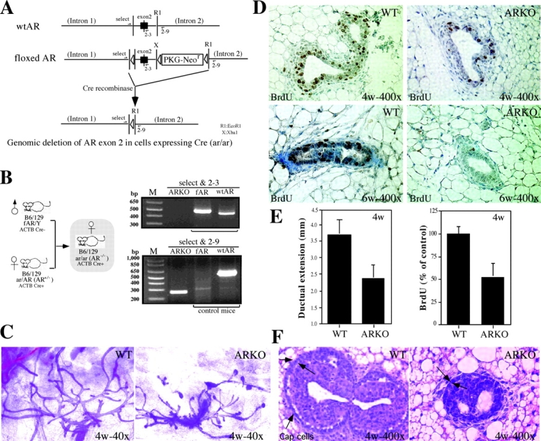
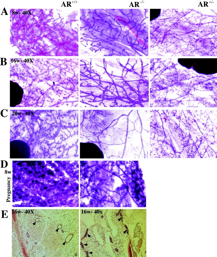
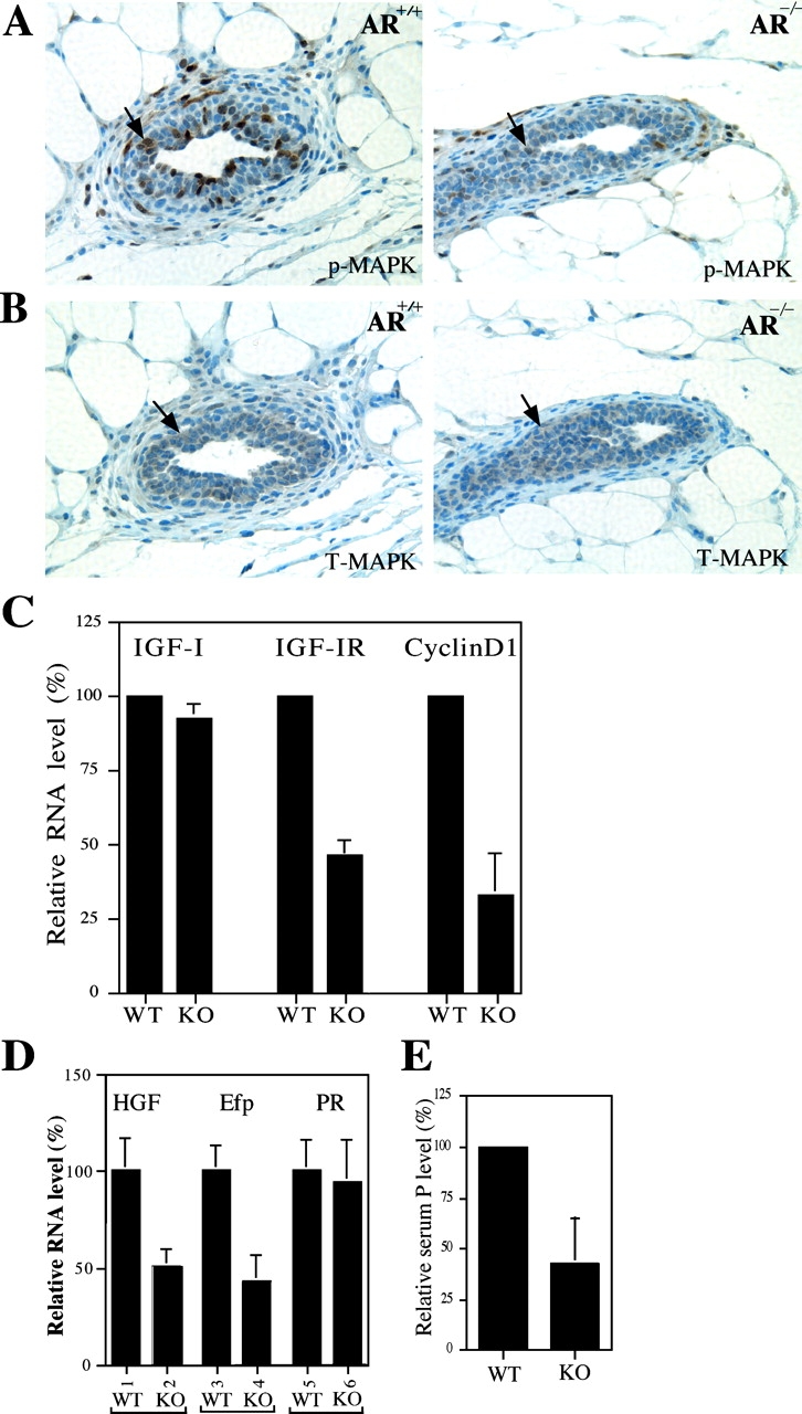
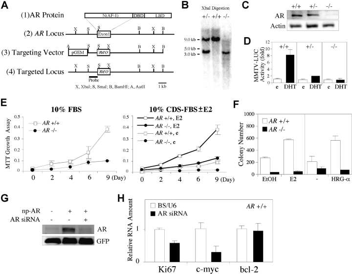
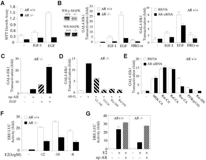
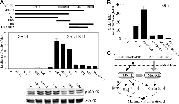
Phenotype analysis of female mice lacking androgen receptor (AR) deficient (AR-/-) indicates that the development of mammary glands is retarded with reduced ductal branching in the prepubertal stages, and fewer Cap cells in the terminal end buds, as well as decreased lobuloalveolar development in adult females, and fewer milk-producing alveoli in the lactating glands. The defective development of AR-/- mammary glands involves the defects of insulin-like growth factor I-insulin-like growth factor I receptor and mitogen-activated protein kinase (MAPK) signals as well as estrogen receptor (ER) activity. Similar growth retardation and defects in growth factor-mediated Ras/Raf/MAPK cascade and ER signaling are also found in AR-/- MCF7 breast cancer cells. The restoration assays show that AR NH2-terminal/DNA-binding domain, but not the ligand-binding domain, is essential for normal MAPK function in MCF7 cells, and an AR mutant (R608K), found in male breast cancer, is associated with the excessive activation of MAPK. Together, our data provide the first in vivo evidence showing that AR-mediated MAPK and ER activation may play important roles for mammary gland development and MCF7 breast cancer cell proliferation.


Figures

Figure 1.
Figure 1.
The generation and characterization of immature female AR −/− mice. (A) Gene targeting strategies. To generate female AR −/− mice, a Cre-lox strategy for conditional knockout was applied. The Cre-lox system uses the expression of P1 phage Cre recombinase to catalyze the excision of DNA located between flanking lox sites. (B) Breeding strategy of female AR −/− mice and genotyping of female AR −/− mice. Using the Cre-lox strategy, the targeted exon 2 of AR is not disrupted but floxed in the male mice. X and RI represent XbaI and EcoRI restriction enzyme sites, respectively. Thus, the AR functions normally in male mice, which can be bred with female AR+/− ACTB Cre+ mice and, thereby, generate homozygous female AR −/− mice. For examining the X chromosome with floxed AR, primers select and 2-3 are used. Select is located in the intron 1 with sequence 5′-GTTGATACCTTAACCTCTGC-3′. 2-3 is the 3′ end primer located in the exon 2 with the sequence 5′-CTTCAGCGGCTCTTTTGAAG-3′. This pair of primers will amplify a product with ∼510 bp for floxed AR and ∼460 bp for wt AR. For examining the AR knockout locus, primers select and 2-9 were used. 2-9 is located in intron 2 with the sequence 5′-CTTACATGTACTGTGAGAGG-3′. The PCR product size from this pair of primers would be ∼270 bp for AR knockout allele and ∼600 bp for wt AR allele. The expression of Cre and internal control IL-2 was also confirmed by PCR genotyping. (C) The defects in the ductal development of mammary gland in immature female AR −/− mice. Whole breast mounts from 4-wk-old female AR −/− mice show lessened extension of mammary ducts, as compared with age-matched AR +/+ mice. (D) The decreases in the percentage of BrdU-positive staining (brown) are observed in both 4- and 6-wk-old AR −/− mice. (E) Statistic analyses of the distance of ductal extension in female AR −/− and AR +/+ mice (left). Statistic analyses of BrdU staining signal in female AR −/− and AR +/+ mice (right, n = 5 for each group). (F) The number of Cap cells (arrows) in TEB of AR −/− mice is less than that in the AR +/+ mice.
Figure 2.
Figure 2.
AR −/− mammary glands show the defects of the terminal branching and alveologenesis during maturity and pregnancy. Whole breast mounts from 8-, 16-, and 20-wk-old mature and 8-wk-old pregnant female mice were examined. (A–C) Less secondary and tertiary terminal branching in AR −/− mice, compared with AR +/+ and AR +/− mice. (C) Early degeneration occurs in AR −/− mammary glands in 20-wk-old mice. (D) The decreased milk-producing lobuloalveolar development in the 8-wk-old pregnant AR −/− mice. (E) Using hematoxylin and eosin staining, the results indicate that the shrunken ductal space occurs in some AR −/− mammary glands in 16–20-wk-old mice (n = 4 for each group).
Figure 3.
Figure 3.
The reduced MAPK activity and mRNA expression of IGF-IR, HGF, and Efp in AR −/− mammary glands. (A) Reduced MAPK activities are observed in AR −/− mammary glands of 4-wk-old mice. Mammary glands were stained with anti–phospho-MAPK (p-MAPK) antibody. The decrease of positive staining (brown) indicates the reduced MAPK activity in AR −/− mammary glands. (B) The similar total MAPK (T-MAPK) staining results are observed in the mammary glands from 4-wk-old AR +/+ and AR −/− mice. Adjacent sections were used for p-MAPK and T-MAPK stainings. (C) The mRNA expression of IGF-IR, but not IGF-I, is reduced in AR −/− mice. Total RNA was extracted from 4-wk-old AR +/+ and AR −/− mice and quantitated by real-time RT-PCR. Cyclin D1, a proliferation indicator, is also reduced in mammary glands of female AR −/− mice. (D) The mRNA expressions of two ER target genes, HGF and Efp, are reduced in AR −/− mice. The expression of PR mRNA has no significant difference (bars 5 vs. 6). Total RNA was extracted from 4-wk-old AR +/+ and AR −/− mice injected with E2 (n = 5 for each group). (E) The serum levels of P were reduced in 12–16-wk-old adult female AR −/− mice.
Figure 4.
Figure 4.
Targeted deletion of AR gene in MCF7 cells results in severe defects in cell proliferation and colony formation. (A) Schematic diagram of the strategy of targeting AR genes in MCF7 cells. (B) Genotyping by Southern blot analysis. Genomic DNA extracted from neomycin-resistant clones was digested with XbaI. The untargeted and targeted loci produced ∼9.0-kb and 3.5-kb bands, respectively. (C) The AR protein was ablated in AR −/− MCF7 cells. (D) The ligand-activated transcriptional activity of AR was reduced in AR +/− MCF7 cells and abrogated in AR −/− MCF7 cells, compared with AR +/+ MCF7 cells. (E) The proliferation of AR −/− MCF7 cells was reduced in media containing 10% FBS (left) or 10% CDS-FBS with ethanol (e) or 10−10 M E2 (right), compared with AR +/+ MCF7 cells, using the MTT proliferation assay. (F) The soft-agar colony formation capacity of AR −/− MCF7 cells was largely reduced compared with AR +/+ MCF7 cells. (G) AR siRNA efficiently knocked down AR in the cells transfected with np-AR. Full length of the 110-kD AR was detected by anti-AR antibody (NH27) using Western blotting assays. Cells were cotransfected with pEGFP-C1 vector for normalization of transfection efficiency. GFP expression was detected with anti-GFP antibody. (H) The mRNA expression of Ki67 and c-myc, but not Bcl-2, was reduced in AR +/+ MCF7 cells transfected with AR siRNA using electroporation, compared with the cells transfected with vector alone. Electroporation was performed using 0.4-cm cuvettes and Gene Pulser II set at 280 V and 950 μF.
Figure 5.
Figure 5.
AR was essential for growth factor and estrogen signaling pathway. (A) Growth factor–induced cell proliferation was impaired in AR −/− MCF7 cells, compared with AR +/+ MCF7 cells. Cultures were incubated with RPMI 1640 media containing 0.2% serum treated with or without growth factors for 8 d. (B) The steady-state level of the active form of MAPK was lower in AR −/− MCF7 cells than that in AR +/+ MCF7 cells, when cells were cultured in the media containing 1% HI-FBS for 5 d (left). Growth factor–induced transcriptional activity of GAL4-Elk1 was diminished in AR −/− MCF7 cells (middle) and in AR +/+ MCF7 cells transfected with AR siRNA (right). (C) The reduced MAPK activity was restored by np-AR, which expresses full-length AR and is driven by natural AR promoter. np-AR synergistically enhanced EGF-induced GAL4-Elk1 transactivation. (D) The AR-FL–activated GAL4-Elk1 transactivation can be inhibited by a MAPK phosphatase (CL-100), a specific inhibitor U0126, dominant-negative Ras (Ras-DN), or Raf (Raf-DN). AR-FL, full-length WT AR. (E) Constitutively activated MEK1 (MEK-CA), Ras (Ras-CA), or Raf (Raf-CA), but not Rac (Rac-CA) or PI3K (p110 catalytic subunit), can block the suppressive effect of AR siRNA on the activity of GAL4-Elk1. (F) The transcriptional activity of ER is reduced in AR −/− MCF7 cells, compared with AR +/+ MCF7 cells. (G) The reduced ER activity in AR −/− MCF7 cells was restored by np-AR. pG5-Luc and ERE-Luc were the reporters for GAL4-Elk1 and ER, respectively. pRL-TK was used as an internal control. Transfections were performed using SuperFect according to the manufacturer's instructions. Values shown are the mean ± SD from at least four independent experiments.
Figure 6.
Figure 6.
The NH2 terminus/DBD of AR were required for normal MAPK activation, and an AR mutant (R608K) was associated with the excessive activation of MAPK. (A) Ectopical expression of AR restored the defective MAPK activity in AR −/− MCF7 cells. The NH2 terminus together with the DBD, but not N, DBD, LBD, or LBD-dH12 alone, were required for activating MAPK, using a transient transfection assay (middle) and a Western blot (bottom) with anti–phospho-MAPK and anti-MAPK antibodies. All of the sequence constructs were FLAG-tagged and inserted into pCDNA3 vector. V, vector alone. dH4–12, AR with deletion from helix 4 to helix 12. (B) The AR-R608K–induced GAL4-Elk1 transactivation was higher than AR-FL. AR-R614H-dprm, containing a point mutation (R614H) and a dprm, lost the ability to activate MAPK, whereas AR-R614H or AR-dprm still partially retained MAPK activation capacity. pG5-Luc was the reporter for GAL4-Elk1, and 5 ng/well pRL-TK was used for internal control. Values shown are mean ± SD from at least four independent experiments. (C) The proposed molecular mechanisms. The AR abrogation in mammary glands or mammary cancer cells retards the growth or development via the impairments of the growth factor and ER signaling pathways. The reduced ER activity, as demonstrated by the decreased target gene expression (Efp and HGF), may partly result from the impairment of the growth factors/MAPK signaling pathway. The reduced PR activity may be due to the reduction of ER activity and/or the serum level of P after puberty but not before puberty (asterisk). The decreased cyclin D1 expression may be caused by the impairments of both the growth factor/MAPK and ER signaling pathways. Together, these impaired signals may contribute to the developmental defects of mouse mammary glands in AR −/− mice and growth inhibition in breast cancer cells lacking AR.

Similar articles

Cited by

References

    1. Chang, C.S., J. Kokontis, and S.T. Liao. 1988. Molecular cloning of human and rat complementary DNA encoding androgen receptors. Science. 240:324–326. - PubMed
    1. McKenna, N.J., and B.W. O'Malley. 2002. Combinatorial control of gene expression by nuclear receptors and coregulators. Cell. 108:465–474. - PubMed
    1. Secreto, G., and B. Zumoff. 1994. Abnormal production of androgens in women with breast cancer. Anticancer Res. 14:2113–2117. - PubMed
    1. Berrino, F., P. Muti, A. Micheli, G. Bolelli, V. Krogh, R. Sciajno, P. Pisani, S. Panico, and G. Secreto. 1996. Serum sex hormone levels after menopause and subsequent breast cancer. J. Natl. Cancer Inst. 88:291–296. - PubMed
    1. Dimitrakakis, C., J. Zhou, and C.A. Bondy. 2002. Androgens and mammary growth and neoplasia. Fertil. Steril. 77(Suppl.):26–33. - PubMed

Publication types