. 2004 Jan 6;101(1):82-6.
doi: 10.1073/pnas.2237114100. Epub 2003 Dec 15.

Loss of poly(ADP-ribose) glycohydrolase causes progressive neurodegeneration in Drosophila melanogaster

Affiliations

Loss of poly(ADP-ribose) glycohydrolase causes progressive neurodegeneration in Drosophila melanogaster

Shuji Hanai et al. Proc Natl Acad Sci U S A. .

Abstract

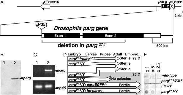
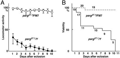
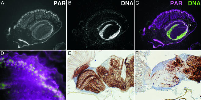
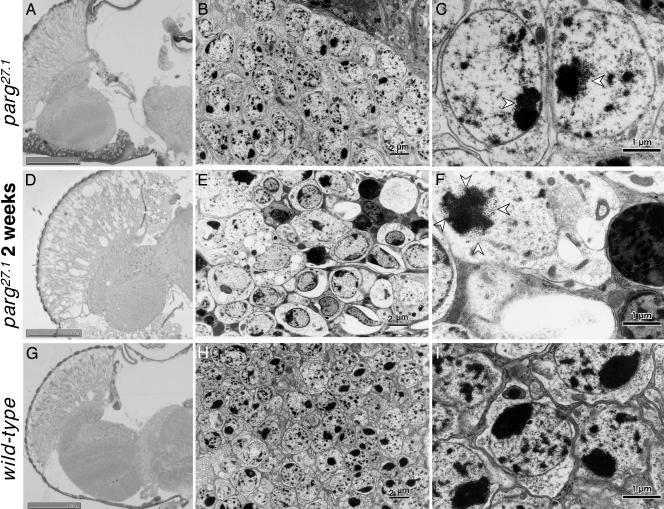
Poly(ADP-ribosyl)ation has been suggested to be involved in regulation of DNA repair, transcription, centrosome duplication, and chromosome stability. However, the regulation of degradation of poly(ADP-ribose) and its significance are not well understood. Here we report a loss-of-function mutant Drosophila with regard to poly(ADP-ribose) glycohydrolase, a major hydrolyzing enzyme of poly(ADP-ribose). The mutant lacks the conserved catalytic domain of poly(ADP-ribose) glycohydrolase, and exhibits lethality in the larval stages at the normal development temperature of 25 degrees C. However, one-fourth of the mutants progress to the adult stage at 29 degrees C but showed progressive neurodegeneration with reduced locomotor activity and a short lifespan. In association with this, extensive accumulation of poly(ADP-ribose) could be detected in the central nervous system. These results suggest that poly(ADP-ribose) metabolism is required for maintenance of the normal function of neuronal cells. The phenotypes observed in the parg mutant might be useful to understand neurodegenerative conditions such as the Alzheimer's and Parkinson's diseases that are caused by abnormal accumulation of substances in nervous tissue.


Figures

Fig. 1.
Fig. 1.
Characterization of the Drosophila parg mutant. (A) The upper line denotes genes in the parg locus predicted by the European Drosophila Genome Sequencing Consortium (GenBank accession no. Z98254). The structure of the Drosophila parg gene is shown in the lower line, and the parg27.1 mutant deletion is indicated by the bracket. Boxes denote exons, and filled boxes denote coding regions. The position of a P element insertion is shown. (B) Genomic Southern blotting of the parg gene. Lane 1, parg27.1/Y; lane 2, wild type. (C) parg transcripts examined by RT-PCR. Lane 1, parg27.1/Y; lane 2, wild-type. rp49 was used as a control. (D) Effects of temperature on development of parg mutants. (E) Relative content of poly(ADP-ribose) examined by ELISA using an anti-poly(ADP-ribose) antibody.
Fig. 2.
Fig. 2.
Locomotor activity and survival of parg27.1/Y and parg27.1/FM7 genotypes. (A) Locomotor activity was measured as described in Materials and Methods. (B) Viability of the same group.
Fig. 3.
Fig. 3.
Accumulation of poly(ADP-ribose) and disruption of axon structures in the parg mutant. (A) Poly(ADP-ribose) in a parg27.1/Y adult as detected with anti-poly(ADP-ribose) antibody. (B) Nuclei are stained with Hoechst dye 33342. A part of the sagittal section of the head is shown. (C) Merging of poly(ADP-ribose) (magenta) and DNA (green). The box in C is shown in D at higher magnification. Axon staining of the horizontal section of adult head at 10 days after eclosion is shown in E for the wild type and in F for parg27.1/Y mutant.
Fig. 4.
Fig. 4.
Microscopic and electron-microscopic analyses of adult brain. Horizontal section through adult heads. (AC) parg27.1/Y at a few days after eclosion. (DF) parg27.1/Y at 2 weeks after eclosion. (GI) Wild type. A, D, and G show light microphotographs of tissue stained with toluidine blue; B, E, and H show electron microphotographs; C, F, and I show higher magnification of parts of B, E, and H, respectively. Open arrowheads indicate granular structures typical in the parg27.1/Y genotype. (Scale bars, 2 μm in B, E, and H and 1 μm in C, F, and I.)

References

    1. de Murcia, G. & Shall, S. (2000) PolyADP-Ribosylation Reactions (Oxford Univ. Press, Oxford).
    1. Bouchard, V. J., Rouleau, M. & Poirier, G. G. (2003) Exp. Hematol. 31, 446–454. - PubMed
    1. Masutani, M., Nakagama, H. & Sugimura, T. (2003) Genes Chromosomes Cancer 38, 339–348. - PubMed
    1. Kraus, W. L. & Lis, J. T. (2003) Cell 113, 677–683. - PubMed
    1. Kanai, M., Tong, W. M., Sugihara, E., Wang, Z. Q., Fukasawa, K. & Miwa, M. (2003) Mol. Cell. Biol. 23, 2451–2462. - PMC - PubMed

Publication types

MeSH terms