. 2003 Dec;112(12):1831-42.
doi: 10.1172/JCI19774.

Superoxide-mediated activation of uncoupling protein 2 causes pancreatic beta cell dysfunction

Affiliations

Superoxide-mediated activation of uncoupling protein 2 causes pancreatic beta cell dysfunction

Stefan Krauss et al. J Clin Invest. 2003 Dec.

Abstract

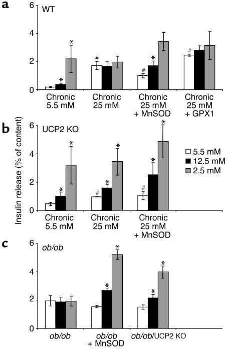
Failure to secrete adequate amounts of insulin in response to increasing concentrations of glucose is an important feature of type 2 diabetes. The mechanism for loss of glucose responsiveness is unknown. Uncoupling protein 2 (UCP2), by virtue of its mitochondrial proton leak activity and consequent negative effect on ATP production, impairs glucose-stimulated insulin secretion. Of interest, it has recently been shown that superoxide, when added to isolated mitochondria, activates UCP2-mediated proton leak. Since obesity and chronic hyperglycemia increase mitochondrial superoxide production, as well as UCP2 expression in pancreatic beta cells, a superoxide-UCP2 pathway could contribute importantly to obesity- and hyperglycemia-induced beta cell dysfunction. This study demonstrates that endogenously produced mitochondrial superoxide activates UCP2-mediated proton leak, thus lowering ATP levels and impairing glucose-stimulated insulin secretion. Furthermore, hyperglycemia- and obesity-induced loss of glucose responsiveness is prevented by reduction of mitochondrial superoxide production or gene knockout of UCP2. Importantly, reduction of superoxide has no beneficial effect in the absence of UCP2, and superoxide levels are increased further in the absence of UCP2, demonstrating that the adverse effects of superoxide on beta cell glucose sensing are caused by activation of UCP2. Therefore, superoxide-mediated activation of UCP2 could play an important role in the pathogenesis of beta cell dysfunction and type 2 diabetes.


Figures

Figure 1
Figure 1
Effects of superoxide on proton conductance of kidney (a and c) and spleen (b and d) mitochondria isolated from WT (a and b) and UCP2-deficient (c and d) mice. Proton leak titration was performed with or without addition of a superoxide-generating system (xanthine plus xanthine oxidase), essentially as described previously (30) and in Methods. Graphs show the rate of proton leak as a function of its driving force, mitochondrial membrane potential. Open squares, control; filled squares, xanthine (50 μM) plus xanthine oxidase (0.2 mU/3.5 ml for kidney mitochondria, 0.1 mU/3.5 ml for spleen mitochondria); open circles, xanthine/xanthine oxidase plus 500 μM GDP. Western blot analysis confirmed that UCP2 protein was present in kidney (e) and spleen mitochondria (8) of WT mice, and that UCP2 protein was absent in mitochondria from UCP2 KO mice. Proton leak data are means ± SEM of three independent experiments.
Figure 2
Figure 2
Effect of MnTBAP on proton leak, mitochondrial membrane potential, ATP, and oxygen consumption in intact thymocytes. (a and b) Proton leak in thymocytes. Thymocytes were incubated with oligomycin to block protons from re-entering the mitochondrial matrix via ATP synthase (top right points of each line). A submaximal concentration (80 nM) of the electron-transport chain inhibitor myxothiazol was added to reduce mitochondrial membrane potential to the resting-cell level (bottom left points in each line). Analyses were performed in thymocytes isolated from WT (a) and UCP2-deficient mice (b). Circles, control; squares, proton leak in the presence of MnTBAP (30 μM). Results are expressed as means ± SEM (n = 4–6). (ce) Mitochondrial membrane potential, total cell ATP, and oxygen consumption were measured in resting thymocytes (no inhibitors added). White bars, control; black bars, 30 μM MnTBAP added. Cells were isolated from WT (left set of bars in each panel) and UCP2-deficient mice (right set of bars in each panel). Results are expressed as means ± SEM (n = 3–7). *P < 0.05, treated vs. untreated control; #P < 0.05, UCP2-deficient control vs. WT control.
Figure 3
Figure 3
Role of superoxide in dispersed islet cells and pancreatic islets in regulating mitochondrial membrane potential (ac), ATP levels (d), and insulin secretion (e). (a) Pseudocolor-coded representative images of TMRM fluorescence intensity in WT and UCP2-deficient dispersed islet cells at the beginning (left) and at the end of the acquisition sequence (right, with 25 μM MnTBAP). After 4 minutes, cells were exposed to 25 μM MnTBAP. (b) Quantitation of the TMRM fluorescence changes over mitochondrial regions. Where indicated by the arrow, 25 μM MnTBAP was added, except for control (WT, no addition). Results are means ± SD (n = 4). (ce) Quantitation of mitochondrial membrane potential, total ATP, and insulin secretion in islets. Islets were isolated from WT and UCP2 KO mice. Islets were treated with MnTBAP, or adenovirus driving the overexpression of MnSOD or GPX1. In some cases, islets were treated with adenovirus driving the expression of GFP as a control for the MnSOD adenovirus studies. In e, insulin content of islets was as follows: WT, 248 ± 11 ng per islet; UCP2 KO, 251 ± 16 ng per islet. These values were used to express insulin secretion as percentage of insulin content. For ce, results are means ± SEM. (c) n = 3–5; (d) n = 8–20 (n = 3 for GFP control); (e) n = 3–5. *P < 0.05, treated vs. untreated control; #P < 0.05, UCP2-deficient control vs. WT control.
Figure 4
Figure 4
UCP2 expression and mitochondrial superoxide production in islets and dispersed islet cells. (a) UCP2 mRNA expression levels in islets cultured for 3 days at low (5.5 mM) and high glucose (25 mM). Expression levels are expressed as relative to the low-glucose (5.5 mM) incubation group. Astrisks indicate P < 0.05, high glucose vs. low glucose control. (b) Mitochondrial superoxide production in low and high glucose in dispersed islet cells from WT and UCP2 KO mice, as measured by time-dependent conversion of the superoxide-sensitive dye hydroethidine (17). Results are means ± SEM (n = 4–7). *P < 0.05, WT (25 mM glucose) vs. WT (5.5 mM glucose); #P < 0.05, UCP2-deficient vs. WT at 5.5 mM glucose; †P < 0.05, UCP2-deficient cells (25 mM glucose) vs. UCP2-deficient cells (5.5 mM glucose). (c) Mitochondrial superoxide production in dispersed islet cells from WT, ob/ob, and ob/ob/UCP2 KO mice, as measured by conversion of the superoxide-sensitive dye hydroethidine. ΔF/min denotes the change in relative fluorescence intensity (F) as a function of time. Results are means ± SEM (n = 3). *P < 0.05, ob/ob or ob/ob/UCP2 KO vs. WT; #P < 0.05, ob/ob/UCP2 KO vs. ob/ob.
Figure 5
Figure 5
Effects of hyperglycemia and obesity on β cell dysfunction in WT and UCP2-deficient islets, with or without overexpression of MnSOD and GPX1. Pancreatic islets were isolated from WT (a) or UCP2 KO mice (b) and subjected to chronic incubations at low and high glucose. In a separate experiment, islets were isolated from ob/ob mice (c). Adenovirus-driven overexpression of MnSOD and GPX1 was used where indicated. Islets were incubated for a total of 72 hours and washed, and then insulin-secretion studies were performed using three different concentrations of glucose (5.5, 12.5, and 25 mM). Insulin- and DNA-content data are reported for reference in Table 1. Results are means ± SEM of three independent experiments (a and b) or five repeats of one representative experiment (c). *P < 0.05, higher glucose vs. lowest glucose condition in each group; #P < 0.05, basal secretion (chronic hyperglycemia) vs. basal secretion (5.5 mM glucose).

Comment in

References

    1. Matthews DR, Clark A. B-cell defects and pancreatic abnormalities in non-insulin-dependent diabetes mellitus. In Textbook of diabetes. JC Pickup and G Williams, editors Blackwell Science Oxford, United Kingdom. 1997;21:21–16.
    1. Poitout V, Robertson RP. Minireview. Secondary beta-cell failure in type 2 diabetes: a convergence of glucotoxicity and lipotoxicity. Endocrinology. 2002;143:339–342. - PubMed
    1. Robertson, R.P., et al. 2000. Glucose toxicity of the beta cell: cellular and molecular mechanisms. In Diabetes mellitus. A fundamental and clinical text. D. Le Roith, S. Taylor, and J.M. Olefsky, editors. Lippincott Williams & Wilkins. Philadelphia, Pennsylvania, USA. 125–132.
    1. Jaburek M, et al. Transport function and regulation of mitochondrial uncoupling proteins 2 and 3. J. Biol. Chem. 1999;274:26003–26007. - PubMed
    1. Echtay KS, Winkler E, Frischmuth K, Klingenberg M. Uncoupling proteins 2 and 3 are highly active H(+) transporters and highly nucleotide sensitive when activated by coenzyme Q (ubiquinone) Proc. Natl. Acad. Sci. U. S. A. 2001;98:1416–1421. - PMC - PubMed

Publication types

MeSH terms