. 2003 Dec;112(12):1843-50.
doi: 10.1172/JCI17450.

Auricular chondritis in NOD.DQ8.Abetao (Ag7-/-) transgenic mice resembles human relapsing polychondritis

Affiliations

Auricular chondritis in NOD.DQ8.Abetao (Ag7-/-) transgenic mice resembles human relapsing polychondritis

Veena Taneja et al. J Clin Invest. 2003 Dec.

Abstract

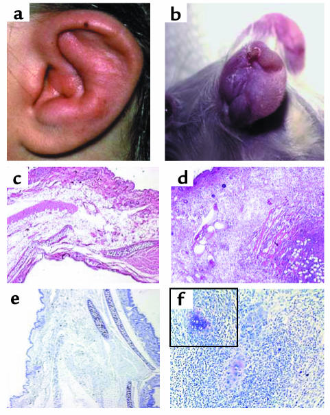
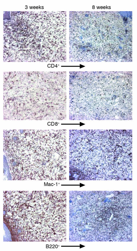
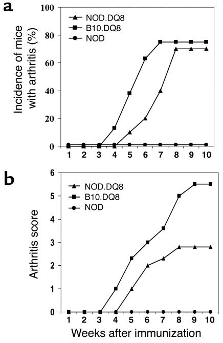
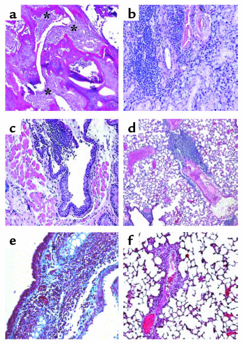
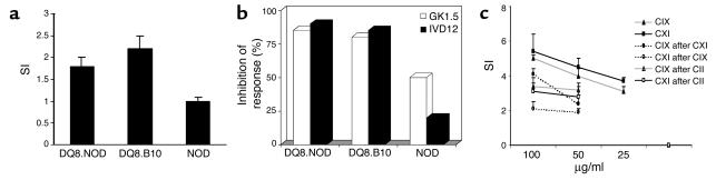
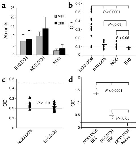
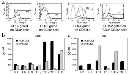
Relapsing polychondritis is a multisystem autoimmune disease involving cartilage destruction but no known causative antigen. HLA-DQ8 has been associated with various autoimmune diseases in humans. To study the role of DQ8 in autoimmune diseases, we have generated transgenic mice expressing DQ8 (DQA1*0301, DQB1*0302) in a NOD background lacking endogenous class II molecules (Abetao). Upon immunization with type II collagen (CII), 85% of NOD.DQ8 mice develop severe experimental polychondritis, auricular chondritis, and polyarthritis, with clinical and histological similarities to relapsing polychondritis (RP) in humans. CII-immunized mice mount a T cell response and produce Ab's to type IX collagen (CIX) and self-CII. Transgene-negative littermates do not develop any serological and clinical manifestations following immunization. B10.DQ8 transgenic mice develop polyarthritis and Ab's to CII only. The susceptibility to auricular chondritis in NOD.DQ8 mice can be attributed to response to CIX. A higher number of activated cells, CD4+CD44(hi)CD62L(lo), and lower regulatory cells CD4+CD152+CD25+ were observed in NOD.DQ8 mice compared with B10.DQ8 mice. The NOD.DQ8 mice provide a model of RP with a high disease incidence and multiple organ involvement to investigate putative autoantigen and regulatory cells involved in disease pathogenesis. An experimental model restricted by the human class II molecule will be valuable when studying the role of various collagens in immunologic and pathologic responses in human RP.


Figures

Figure 1
Figure 1
Swelling and edema with a shrunken ear as observed in a human ear (a) and an affected mouse (b) with auricular chondritis. H&E-stained section of a normal (c) and a chondritic ear (d) of NOD.DQ8 transgenic mice showing massive infiltration of cells in the chondritic ear. Toludine blue staining of a normal ear (e) and a chondritic ear (f) showing destruction of cartilage in the latter, with residual cartilage seen among infiltrates as shown within the boxed area (magnification ×200). Micrographs are ×50 magnification.
Figure 2
Figure 2
Characterization of the infiltrating cells in chondritic ear of NOD.DQ8 mice at 3 weeks and 8 weeks after onset of disease. At 3 weeks, CD4+, B220+, and Mac-1+ cells were seen in abundance distributed throughout the section. Eight weeks after onset of chondritis, infiltrating CD4+, CD8+, and Mac-1+ cells were reduced. B cells were present in aggregates, giving an appearance of follicles.
Figure 3
Figure 3
(a) Incidence and onset of arthritis after immunization with CII showed a delayed onset of arthritis (P < 0.01) with similar incidence in NOD.DQ8 mice compared with B10.DQ8 mice. (b) NOD.DQ8 mice developed milder arthritis compared with B10.DQ8 mice (P < 0.01).
Figure 4
Figure 4
Sections from various organs of NOD.DQ8 mice stained with H&E. (a) Representative inflammatory arthritis of the knee from a NOD.DQ8 mouse. Significant mononuclear infiltration with cartilage destruction and pannus formation is seen (*). (b) Salivary glands showing mononuclear infiltrate suggestive of sialadentitis. (c) Infiltration with mononuclear cells in the trachea. (d) A section of lung shows perivascular infiltration in lungs. Accustain trichrome staining of frozen sections of trachea (e) and lungs (f). Only tracheas showed deposition of collagen, as seen by the blue color. Micrographs ad are at ×100 magnification; e and f are at ×200 magnification.
Figure 5
Figure 5
(a) In vitro response of lymph node cells (LNCs) to chick CII (50 μg/ml) from primed mice in transgenic and control mice. The results shown are mean ± SD of three experiments. SDs were lower than 10% in all experiments. (b) Inhibition of response was measured from LNCs cultured as above in the presence of specific Ab’s, DQ (IVD12) or mCD4 (GK1.5). The results are shown as the percentage of inhibition of response by a specific Ab. (c) NOD.DQ8 mice primed with CIX and CXI mount a dose-dependent response to a specific collagen. Cross-reactive response to CIX was observed in CII-immunized mice. The data is mean ± SD of all experiments. SI, stimulation index.
Figure 6
Figure 6
NOD.DQ8 mice produce higher levels of Ab’s to self, MsII, and ChII than B10.DQ8 mice (anti-MsII: P < 0.001; anti-ChII: P < 0.05). Bars depict mean ± SD (units per milliliter) of anti-collagen Ab’s. NOD mice produced very low levels of anti-CII and anti-MsII Ab’s compared with transgenic mice (P < 0.001). (b) Only NOD.DQ8 mice produced significant levels of anti-CIX Ab’s following immunization with CII. Aβo mice were used as negative controls and are depicted as cut-off line. (c) IgM-RF was produced at higher levels in NOD.DQ8 compared with B10.DQ8 mice (P < 0.01). Sera from MRL/lpr and B6 mice were used as positive (dotted line) and negative (solid line) controls, respectively. Horizontal bar for each strain indicates mean values of IgM-RF. (d) CIX-primed NOD.DQ8 mice produce significantly high amounts of anti-CIX Ab’s compared with B10.DQ8 mice. Aβo mice, which were used as negative controls, are depicted as cut-off line. BIX, bovine CIX.
Figure 7
Figure 7
(a) Cell surface markers on splenic cells as determined by FACS after labeling them with specific mAb’s in B10.DQ8 mice (broken line) and NOD.DQ8 mice (solid line). NOD.DQ8 mice had a lower number of CD8+CD25hi, CD152+CD4+CD25+, and a higher number of B220+CD25+ cells than B10.DQ8 mice. The data shown were obtained from spleen cells pooled from two mice per strain and represent at least two separate assays. (b) NOD.DQ8 mice produce high levels of TGF-β. Cytokines produced by splenic cells isolated from primed mice and cultured in vitro in the presence of CII and (c) CIX. For each experiment, cells were pooled from two mice per strain.

References

    1. Zeuner M, Straub RH, Albert ED, Scholmerich J, Lang B. Relapsing polychondritis: clinical and immunogenetic analysis of 62 patients. J. Rheumatol. 1997;24:96–101. - PubMed
    1. McAdam LP, O’Hanlan MA, Bluestone R, Pearson CM. Relapsing polychondritis: perspective study of 23 patients and a review of literature. Medicine. 1976;55:193–215. - PubMed
    1. Issak BL, Liesegang TJ, Michet CJ. Ocular and systemic findings in relapsing polychondritis. Opthalmology. 1986;93:681–689. - PubMed
    1. Homma S, Matsumoto T, Abe H, Fukuda Y, Suzuki M. Relapsing polychondritis. Pathological and immunological findings in an autopsy case. Acta Pathol. Jpn. 1984;34:1137–1146. - PubMed
    1. Damiani JM, Levine HL. Relapsing polychondritis: report of ten cases. Laryngoscope. 1979;89:929–946. - PubMed

Publication types

MeSH terms