. 2003 Dec;112(12):1906-14.
doi: 10.1172/JCI19826.

Renal Ca2+ wasting, hyperabsorption, and reduced bone thickness in mice lacking TRPV5

Affiliations

Renal Ca2+ wasting, hyperabsorption, and reduced bone thickness in mice lacking TRPV5

Joost G J Hoenderop et al. J Clin Invest. 2003 Dec.

Abstract

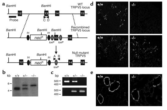
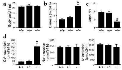
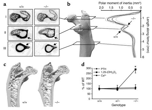
Ca2+ ions play a fundamental role in many cellular processes, and the extracellular concentration of Ca2+ is kept under strict control to allow the proper physiological functions to take place. The kidney, small intestine, and bone determine the Ca2+ flux to the extracellular Ca2+ pool in a concerted fashion. Transient receptor potential (TRP) cation channel subfamily V, members 5 and 6 (TRPV5 and TRPV6) have recently been postulated to be the molecular gatekeepers facilitating Ca2+ influx in these tissues and are members of the TRP family, which mediates diverse biological effects ranging from pain perception to male aggression. Genetic ablation of TRPV5 in the mouse allowed us to investigate the function of this novel Ca2+ channel in maintaining the Ca2+ balance. Here, we demonstrate that mice lacking TRPV5 display diminished active Ca2+ reabsorption despite enhanced vitamin D levels, causing severe hypercalciuria. In vivo micropuncture experiments demonstrated that Ca2+ reabsorption was malfunctioning within the early part of the distal convolution, exactly where TRPV5 is localized. In addition, compensatory hyperabsorption of dietary Ca2+ was measured in TRPV5 knockout mice. Furthermore, the knockout mice exhibited significant disturbances in bone structure, including reduced trabecular and cortical bone thickness. These data demonstrate the key function of TRPV5 in active Ca2+ reabsorption and its essential role in the Ca2+ homeostasis.


Figures

Figure 1
Figure 1
Creation of null mutant mice for the TRPV5 gene locus. (a) Targeted inactivation of the TRPV5 gene. Top, exon 13, encoding the pore-forming unit of TRPV5, is flanked by loxP sites. Middle, recombined TRPV5loxneo locus. Bottom, targeted allele in which exon 13 is deleted by Cre recombinase. Black boxes indicate exons; probe and genotype primers (A–D) are depicted. neoR, neomycin resistance cassette. (b) Representative Southern blot analysis of BamHI-digested tail-derived DNA isolated from TRPV5+/+, TRPV5+/–, and TRPV5–/– mice. (c) Identification of mouse genotype by PCR analysis of tail-derived DNA. The PCR product in the upper gel shows the presence of the wild-type allele (+/+) using primers C and D; the lower gel shows the knockout allele (–/–) using primers A and B. Both alleles are detected in heterozygous animals (+/–). (d) TRPV5 (top images) and kallikrein (lower images) immunohistochemical costaining of kidney cortex from TRPV5+/+ and TRPV5–/– mice. (e) Immunopositive TRPV6 staining of renal distal tubules from TRPV5+/+ and TRPV5–/– mice. A color version of this figure is available online as Supplementary Figure 1 (http://www.jci.org/cgi/content/full/112/12/1906/DC1).
Figure 2
Figure 2
Phenotypic characterization of TRPV5 knockout mice. (a) Body weights at an age of 8 weeks of TRPV5+/+, TRPV5+/–, and TRPV5–/– mice (n = 29–43 mice). (b) The 24-hour urine volumes obtained from mice maintained in metabolic cages (n = 9 mice, three mice per cage). (c) Analysis of pH of urine obtained from TRPV5+/+, TRPV5+/–, and TRPV5–/– mice (n = 9 mice, three mice per cage). (d) Total excretion of Ca2+, Na+, and K+ in TRPV5+/+, TRPV5+/–, and TRPV5–/– mice (n = 9 mice, three mice per cage). Of note, TRPV5+/– mice were similar to TRPV5+/+ mice for the parameters measured. Data are averaged values ± SEM from mice at an age of 8 weeks. *P < 0.05, significant difference from TRPV5+/+ mice.
Figure 3
Figure 3
Renal and duodenal Ca2+ transport assays. (a) Fractional Ca2+ delivery to the LPT, to the distal convolution (DC), and to urine (U), measured by micropuncture experiments in TRPV5+/+ and TRPV5–/– mice. (b) Relation between K+ concentration in tubular fluid of the distal convolution and fractional Ca2+ delivery to these sites. Low K+ concentrations indicate early puncture sites and high K+ concentrations late aspects of distal convolution (2). Values are means ± SEM of six mice per group for urine data and of 19–24 nephrons per group for LPT and distal convolution collections. *P < 0.05 versus TRPV5+/+. (c) Changes in serum Ca2+ (ΔμM) within 10 minutes of administration of 45Ca2+ by oral gavage in TRPV5+/+ and TRPV5–/– mice (n = 12). Data are averaged values ± SEM from mice 8 weeks old. *P < 0.05, significant difference from wild-type mice. (d) Expression of calbindin-D28K and NCX1 mRNA in kidney cortexes of TRPV5+/+ and TRPV5–/– mice (n = 9), assessed by quantitative real-time PCR analysis and calculated as a ratio to HPRT RNA. (e) Expression of TRPV6 and calbindin-D9K mRNA in duodenums of TRPV5+/+ and TRPV5–/– mice, assessed by quantitative real-time PCR analysis and calculated as a ratio to the HPRT RNA (n = 9).
Figure 4
Figure 4
Bone phenotypes of female and male TRPV5+/+ and TRPV5–/– mice. (a) Representative cross-sectional x-ray images of the femoral head (I), the lesser trochanter (II), and the diaphysis (III) in TRPV5+/+ and TRPV5–/– mice. Note the decreased cortical bone width in the trochanter and diaphysis (arrows). (b) MOI as a parameter of bone quality. (c) Three-dimensional reconstruction of femurs from TRPV5+/+ and TRPV5–/– mice. Note the reduced cortical and Tb.Th in TRPV5–/– mice. (d) Changes in serum levels of 1,25-(OH)2D3, PTH, and Ca2+ in TRPV5+/+, TRPV5+/–, and TRPV5–/– mice. Values are expressed as percentage of TRPV5+/+ values ± SEM. The absolute values for PTH are TRPV5+/+, 55 ± 15 pg/ml; TRPV5+/–, 59 ± 14 pg/ml; and TRPV5–/–, 60 ± 16 pg/ml; for 1,25-(OH)2D3, TRPV5+/+, 239 ± 38 pmol/l; TRPV5+/–, 225 ± 32 pmol/l; TRPV5–/–, 691 ± 88 pmol/l; and for free Ca2+ concentration, TRPV5+/+, 1.20 ± 0.02 mM; TRPV5+/–, 1.25 ± 0.03 mM; and TRPV5–/–, 1.20 ± 0.03 mM. *P < 0.05, significant difference from wild-type mice.

References

    1. Hoenderop JG, et al. Molecular identification of the apical Ca2+ channel in 1, 25-dihydroxyvitamin D3-responsive epithelia. J. Biol. Chem. 1999;274:8375–8378. - PubMed
    1. Peng JB, et al. Molecular cloning and characterization of a channel-like transporter mediating intestinal calcium absorption. J. Biol. Chem. 1999;274:22739–22746. - PubMed
    1. Montell C, Birnbaumer L, Flockerzi V. The TRP channels, a remarkably functional family. Cell. 2002;108:595–598. - PubMed
    1. Hoenderop JG, Nilius B, Bindels RJ. Molecular mechanisms of active Ca2+ reabsorption in the distal nephron. Ann. Rev. Physiol. 2002;64:529–549. - PubMed
    1. Yue L, Peng JB, Hediger MA, Clapham DE. CaT1 manifests the pore properties of the calcium-release-activated calcium channel. Nature. 2001;410:705–709. - PubMed

Publication types

MeSH terms