. 2004 Jan 26;117(Pt 3):397-405.
doi: 10.1242/jcs.00868. Epub 2003 Dec 16.

Electrical stimulation directly induces pre-angiogenic responses in vascular endothelial cells by signaling through VEGF receptors

Affiliations

Electrical stimulation directly induces pre-angiogenic responses in vascular endothelial cells by signaling through VEGF receptors

Min Zhao et al. J Cell Sci. .

Abstract

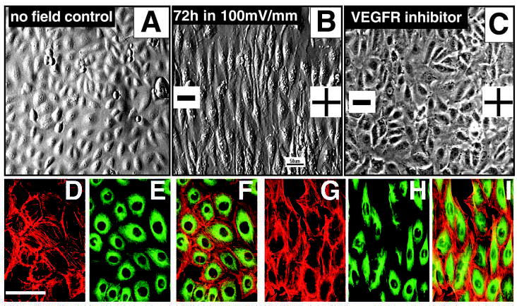
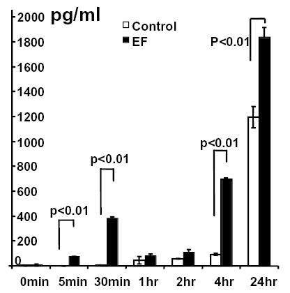
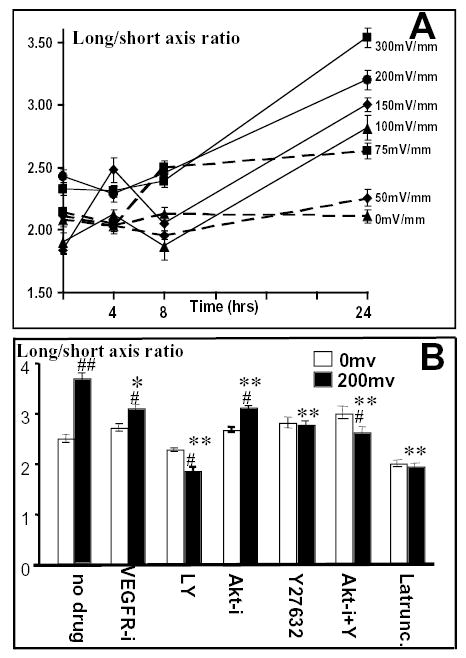
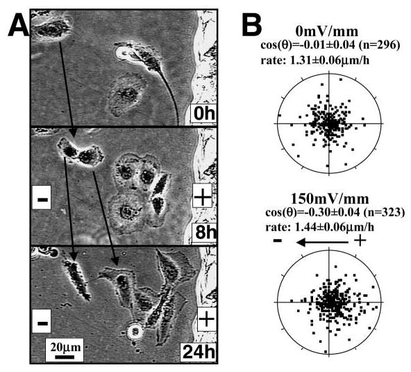
Controlling angiogenesis is crucial. Growth factors and cytokines are key regulators but a full understanding remains elusive. Endogenous electrical potential differences exist within and around the vasculature, both in relation to blood flow and in situations where active angiogenesis occurs, such as wound healing, development and tumor growth. Recent work shows that electrical stimulation induces significant angiogenesis in vivo, through enhanced vascular endothelial growth factor (VEGF) production by muscle cells. We report that applied electric fields (EFs) of small physiological magnitude directly stimulate VEGF production by endothelial cells in culture without the presence of any other cell type. EFs as low as 75-100 mV mm-1 (1.5-2.0 mV across an endothelial cell) directed the reorientation, elongation and migration of endothelial cells in culture. These pre-angiogenic responses required VEGF receptor activation and were mediated through PI3K-Akt and Rho-ROCK signaling pathways, resulting in reorganization of the actin cytoskeleton. This indicates that endogenous EFs might play a role in angiogenesis in vivo by stimulating the VEGF receptor signaling pathway, to induce key pre-angiogenic responses. In addition, it raises the feasibility of using applied EFs to initiate and guide angiogenesis through direct effects on endothelial cells.


Figures

Fig. 1
Fig. 1
Perpendicular orientation and elongation of endothelial cells in a small physiological EF. (A) Control HUVEC cells cultured in the same chamber without EFs showed a typical cobblestone morphology and random orientation. (B) Cells exposed to small applied EFs showed dramatic elongation and perpendicular orientation in the EF. (C) Cells treated with a VEGFR inhibitor that completely abolished perpendicular orientation and significantly inhibited elongation in an applied EF (72 hours, 100 mV mm−1). (G-I) Most actin filaments (red) and microtubules (green) became aligned along the long axis of the cells (12 hours at 150 mV mm−1). (D-F) No-field controls showed no obvious alignment and cell elongation. (A,B) Images taken with Hoffman modulation optics. (C) Image taken with phase-contrast optics. (F,I) Merged images.
Fig. 2
Fig. 2
Endothelial cells reoriented, elongated and migrated directionally in a small physiological EF. Obvious elongation and orientation of cells can be seen after 8 hours in an EF. Directional lamellipodial extension and cell migration were evident at 12 hours and 24 hours after EF exposure. Scratches made on the culture dish as static reference points can be seen to the left and top of each frame.
Fig. 3
Fig. 3
Time and voltage dependency of orientation of endothelial cells in a small physiological EF. (A) Orientation index as a function of time and voltage (n=65–598 from at least two independent experiments). (B) Effects of various drugs on EF-induced perpendicular orientation. Inhibition of PI3K (LY), Akt (Akt-i), Rho (Y27632) and actin polymerization (Latrunc) significantly decreased orientation responses, whereas inhibition of VEGFR (VEGFR-i), or combined inhibition of both Akt and Rho (Akt-i + Y) completely abolished the orientation response. VEGFR-i, VEGFR inhibitor (50 μM); LY, PI3K inhibitor LY294002 (50 μM); Akt-i, Akt inhibitor (50 μM); Y27632, Rho inhibitor (50 μM); Akt-i + Y27632, Akt and Rho inhibitors (10 μM each); Latrunc, Latrunculin (50 μM). Endothelial cells were subjected to EFs of 200 mV mm−1 for 24 hours. n=47–343 from at least two independent experiments. **, P<0.0001 compared with cells exposed to 200 mV mm−1 without drug treatment.
Fig. 4
Fig. 4
Exposure to a physiological EF increased VEGF release from endothelial cells. HUVEC cells were cultured in serum-free DMEM and exposed to an EF of 200 mV mm−1. VEGF in the medium was quantified by ELISA. Values are means±s.e.m.
Fig. 5
Fig. 5
Time and voltage dependency of elongation of endothelial cells in a small physiological EF. (A) Cell elongation as a function of time and voltage (n=65–598 from at least two independent experiments). (B) Effects of various drugs on EF-induced cell elongation. Inhibition of PI3K (LY), Akt (Akt-i), Rho (Y27632) and actin polymerization (Latrunc) significantly decreased the elongation response, as did inhibition of VEGR (VEGFR-i), and the combined inhibition of both Akt and Rho (Akt-i + Y). VEGFR-i, VEGFR inhibitor (50 μM); LY, PI3K inhibitor LY294002 (50 μM); Akt-i, Akt inhibitor (50 μM); Y27632, Rho inhibitor (50 μM); Akt-i + Y27632, Akt and Rho inhibitors (10 μM each); Latrunc, Latrunculin (50 μM). Cells were subjected to EFs of 200 mV mm−1 for 24 hours. n=47–343 from more than two independent experiments. *, P<0.002 compared with cells exposed to 200 mV mm−1 without drug treatment; **, P<0.0001 compared with cells exposed to 200 mV mm−1 without drug treatment; #, P<0.001 compared with same drug treatment but not exposed to EFs; ##, P<0.0001 compared with same drug treatment but not exposed to EFs.
Fig. 6
Fig. 6
Directional endothelial cell migration in a small physiological EF. (A) Endothelial cells in culture exposed to an EF of 100 mV mm−1 migrated directionally towards the anode. Cells migrated slowly but steadily toward the anode over 24 hours. Movement is evident using the static scratch on the culture dish as a reference (right margin). Notice that lamellipodia extended preferentially toward the anode. (B) Scatter plot showing biased migration of endothelial cells in an EF. Cells started at the origin and each dot represents the position of each cell 4 hours later. The distribution of the cells shifted towards the anode. Radius=7 μm.

References

    1. Albuquerque ML, Waters CM, Savla U, Schnaper HW, Flozak AS. Shear stress enhances human endothelial cell wound closure in vitro. Am J Physiol Heart Circ Physiol. 2000;279:H293–H302. - PubMed
    1. Barker AT, Jaffe LF, Vanable JW., Jr The glabrous epidermis of cavies contains a powerful battery. Am J Physiol. 1982;242:R358–R366. - PubMed
    1. Borgens RB, Shi R. Uncoupling histogenesis from morphogenesis in the vertebrate embryo by collapse of the transneural tube potential. Dev Dyn. 1995;203:456–467. - PubMed
    1. Brent TP, Forrester JA. Changes in surface charge of HeLa cells during the cell cycle. Nature. 1967;215:92–93. - PubMed
    1. Burger PC, Chandler DB, Klintworth GK. Corneal neovascularization as studied by scanning electron microscopy of vascular casts. Lab Invest. 1983;48:169–180. - PubMed

Publication types

MeSH terms

Substances