. 2003 Dec 15;17(24):3062-74.
doi: 10.1101/gad.1157503. Epub 2003 Dec 17.

SCFbeta-TRCP links Chk1 signaling to degradation of the Cdc25A protein phosphatase

Affiliations

SCFbeta-TRCP links Chk1 signaling to degradation of the Cdc25A protein phosphatase

Jianping Jin et al. Genes Dev. .

Abstract

Eukaryotic cells respond to DNA damage and stalled replication forks by activating protein kinase-mediated signaling pathways that promote cell cycle arrest and DNA repair. A central target of the cell cycle arrest program is the Cdc25A protein phosphatase. Cdc25A is required for S-phase entry and dephosphorylates tyrosine-15 phosphorylated Cdk1 (Cdc2) and Cdk2, positive regulators of cell division. Cdc25A is unstable during S-phase and is degraded through the ubiquitin-proteasome pathway, but its turnover is enhanced in response to DNA damage. Although basal and DNA-damage-induced turnover depends on the ATM-Chk2 and ATR-Chk1 pathways, how these kinases engage the ubiquitin ligase machinery is unknown. Here, we demonstrate a requirement for SCFbeta-TRCP in Cdc25A turnover during an unperturbed cell cycle and in response to DNA damage. Depletion of beta-TRCP stabilizes Cdc25A, leading to hyperactive Cdk2 activity. SCFbeta-TRCP promotes Chk1-dependent Cdc25A ubiquitination in vitro, and this involves serine 76, a known Chk1 phosphorylation site. However, recognition of Cdc25A by beta-TRCP occurs via a noncanonical phosphodegron in Cdc25A containing phosphoserine 79 and phosphoserine 82, sites that are not targeted by Chk1. These data indicate that Cdc25A turnover is more complex than previously appreciated and suggest roles for an additional kinase(s) in Chk1-dependent Cdc25A turnover.


Figures
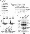
Figure 1.
Figure 1.
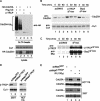
In vivo association of Cdc25A with the SCFβ-TRCP ubiquitin ligase. (A) Disruption of the Cul1 ubiquitin ligase pathway leads to accumulation of Cdc25A. 293T cells were transfected with vectors expressing an N-terminal fragment of Cul1 (residues 1-452; Cul1DN) or empty vector (3 μg) and after 48 h, lysates were subjected to immunoblotting with the indicated antibodies. (B) Disruption of the Cul1 pathway blocks DNA-damage-dependent elimination of Cdc25A. 293T cells were transfected with 3 μg of pCMV-Cul1DN or empty vector subjected to ionizing radiation (10 Gy) prior to analysis of total Cdc25A by immunoblotting. (C) Effect of Cul1DN on cell cycle control. 293T cells were collected 48 h after transfection with either pcDNA3 or pcDNA3-Cul1DN as described in B and then subjected to flow cytometry after staining with propidium iodide. (D) Cdc25A associates with endogenous Cul1 and β-TRCP in the presence and absence of DNA damage. 293T cells were treated with IR (10 Gy). Then, 30 min later, cells were lysed and Cdc25A immune complexes were prepared from 1 mg of extract prior to immunoblotting for Cul1. The blot was stripped and reprobed for β-TRCP. The relative levels of Cdc25A, Cul1, and β-TRCP were determined by densitometry. After normalizing for a small (17%) increase in the amount of Cdc25A in the presence of damage, we determined a 1.85- and 2.5-fold increase in Cdc25A-associated β-TRCP and Cul1 levels in response to DNA damage. (E) Cdc25A associates specifically with the neddylated and activated form of Cul1. Immune complexes were prepared as in D and subjected to electrophoresis together with crude extract (25 μg) to visualize the position of neddylated and unneddylated Cul1. As expected, neddylated Cul1 represents ∼5% of the total Cul1 present in crude extracts.
Figure 2.
Figure 2.
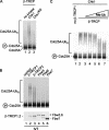
Screening a panel of F-box proteins for association with Cdc25A reveals specific association with β-TRCP1 and β-TRCP2. Vectors expressing the indicated Myc-tagged F-box proteins (1 μg) were cotransfected into 293T cells (2-cm dish) with either pCMV-GST or pCMV-GST-Cdc25A (1 μg) and association with GST-Cdc25A determined 48 h later after purification of GSH-Sepharose. A band cross reacting with anti-Myc antibodies in crude extracts is indicated by the asterisk.
Figure 3.
Figure 3.
Linkage of β-TRCP to elimination of Cdc25A in the presence and absence of DNA damage. (A) Expression of β-TRCP promotes Cdc25A ubiquitination in vivo. 293T cells in 6-cm dishes were transfected with vectors expressing HACdc25A (2 μg), β-TRCP1 (1 μg), and/or His6-Ub (4 μg). After 36 h, cells were lysed in buffers containing 6 M guanidinium-HCl and His6-Ub tagged proteins purified on Ni-NTA beads. Proteins were separated by SDS-PAGE and immunoblotted with anti-HA antibodies. Cells from a parallel transfection were lysed using conventional NP-40-containing buffers (see Materials and Methods) and extracts subjected to immunoblotting (lower panels) using anti-HA to determine the total abundance of HA-Cdc25A and anti-Cul1 as a loading control. (B,C) Accumulation of Cdc25A after DNA damage in the presence of dominant-negative β-TRCP1. 293T cells in 10-cm dishes were transfected with 5 μg of pCMV, pCMV-β-TRCPΔF-box, and pCMV-Skp2ΔF-box. After 48 h, cells were subjected to IR (10 Gy) and lysates were examined by immunoblotting (panel B) at the indicated times with anti-Cdc25A and anti-Cul1 antibodies as a loading control. For analysis of Cdc25A, extracts were subjected to a modified SDS-PAGE procedure that provides larger separation of phosphoryated forms of Cdc25A seen previously (Zhao et al. 2002). The expression of β-TRCPΔF-box and Skp2ΔF-box was confirmed by immunoblotting (panel C). (D,E) Knock-down of β-TRCP by shRNA blocks constitutive and DNA-damage-dependent elimination of Cdc25A. The indicated cells were infected with a single retroviruses expressing shRNA targeting both β-TRCP1 and β-TRCP2 as well a retrovirus expressing shRNA against GFP as a negative control. Then, 48 h later, cells were lysed prior to analysis of β-TRCP and Cul1 levels by immunoblotting (panel D; see Materials and Methods). (E) shRNA-expressing cells were either subjected to ionizing radiation (10 Gy) or left untreated. Cells were lysed 30 min later and 25 μg of extract was subjected to immunoblotting with anti-Cdc25A antibodies. Blots were reprobed with anti-Cdc25C as a loading control.
Figure 4.
Figure 4.
Stabilization of Cdc25A by depletion of β-TRCP in the presence or absence of DNA damage leads to deregulated cyclin E/Cdk2 activity. (A,B) Turnover of Cdc25A requires β-TRCP. 293T cells were transfected with pSUPER-shRNAGFP or pSUPER-shRNAβ-TRCP using lipofectamine 48 h after dual transfection and then either left untreated or subjected to DNA damage (10 Gy). Translation was immediately blocked by addition of cyclohexamide, and cells lysed at the indicated time points prior to SDS-PAGE. (A) Blots were probed with anti-Cdc25A, stripped, and reprobed with anti-Cul1 antibodies. (B) Blots of comparable intensity for shRNAGFP and shRNAβ-TRCP were quantified by densitometry. (C) Depletion of β-TRCP does not alter cell cycle progression. Cells from A were subjected to flow cytometry after staining with propidium iodide. (D) Increased cyclin E/Cdk2 kinase activity in cells depleted of β-TRCP. 293T cells were transfected with vectors expressing shRNA against GFP as control or β-TRCP using the dual-transfection protocol. After 48 h, cells were lysed and cyclin E immune complexes assayed for activity using histone H1 as a substrate. Cyclin E immune complexes were generated using a rabbit polyclonal antibody (C-19 from Santa Cruz Biotechnology). Parallel immunoblots were probed for Cdk2 and cyclin E to demonstrate equal loading. The cyclin E immunoblot was probed with a monoclonal antibody (HE12, Santa Cruz Biotechnology). Controls demonstrated a dramatic accumulation of Cdc25A in response to depletion of β-TRCP whereas Cul1 levels remained unchanged.
Figure 5.
Figure 5.
Chk1-dependent ubiquitination of Cdc25A by SCFβ-TRCP in vitro. (A) Activation of Cdc25A ubiquitination by SCFβ-TRCP requires active Chk1. In vitro translated and 35S-methionine-labeled Cdc25A was subjected to ubiquitination of SCFβ-TRCP complexes assembled in reticulocyte extracts in the absence of kinase or in the presence of active or inactive Chk1 (100 ng) made in insect cells as described in Materials and Methods. Phosphorylation of Cdc25A by Chk1 leads to a small decrease in electrophoretic mobility, as indicated. (B) Both β-TRCP1 and β-TRCP2, but not other WD40-containing F-box proteins, can ubiquitinate Cdc25A in vitro. Assays were performed as in A in the presence of Chk1 with the indicated in vitro translated F-box proteins. (C) Polyubiquitination of Cdc25A by SCFβ-TRCP is attenuated in the presence of methyl ubiquitin. Chk1-dependent Cdc25A ubiquitination reactions were performed in the presence of constant amounts of SCFβ-TRCP and increasing concentrations of methyl ubiquitin. The methyl ubiquitin concentrations used were 0.25, 1, 2.5, 25, and 100 μg/mL.
Figure 6.
Figure 6.
Chk1 phosphorylation sites in Cdc25A are required for SCFβ-TRCP-mediated ubiquitination but do not appear to constitute the major Cdc25A phosphodegron. (A) Schematic representation of Chk1 phosphorylation sites in Cdc25A and comparison of a putative phosphodegron in Cdc25A with the IκBα, β-catenin, and Emi1 phosphodegron recognized by β-TRCP. The Cdc25A residues are designated based on GenBank accession number AAH18642. (B) Identification of residues in Cdc25A important for Chk1-dependent ubiquitination. The indicated Cdc25A mutants were used in SCFβ-TRCP-driven ubiquitination reactions in the presence of Chk1. (C) Arg 474 in β-TRCP1 is required for Chk1-dependent Cdc25A ubiquitination. Ubiquitination of Chk1-phosphorylated Cdc25A was performed in the presence of β-TRCP1 or an R474A mutant. An aliquot of each β-TRCP synthesis reaction was supplemented with 35S-methionine to demonstrate equal expression of β-TRCP proteins (lower panel). (D) Three-dimensional structure of β-TRCP bound to the phosphodegron of β-catenin depicting the interaction of D32 and phosphoserine-33 (pS33) in the phosphodegron with R474 and R285 in β-TRCP. Graphics were generated using Pymol.
Figure 7.
Figure 7.
Involvement of a novel phosphodegron in Cdc25A centered at S82 in association with β-TRCP in vivo. (A,B) A Cdc25A-derived phosphodegron containing at phospho-S79 and phospho-S82 binds β-TRCP in vitro. The indicated synthetic peptides spanning S76-S82 in Cdc25A in phosphorylated or unphosphorylated forms were immobilized on agarose beads and used in binding reactions with in vitro translated and 35S-methionine-labeled β-TRCP1. Bound proteins were separated by SDS-PAGE and visualized by autoradiography. Peptides in A were coupled to Sulfo-link agarose through an N-terminal cysteine while peptides in B were coupled to Affigel-10 through the side chain of the N-terminal lysine. (C) Cdc25A-derived phosphodegrons inhibit ubiquitination of Cdc25A by SCFβ-TRCP in vitro. Assays were performed as described in Figure 5 in the presence of the indicated peptides at 2, 10, 20, and 40 μg/mL. (D) Cdc25A ubiquitination is blocked by an IκBα phosphodegron. Phosphorylated or unphosphorylated synthetic peptides encompassing the IκBα phosphodegron were used in competition reactions with Chk1-dependent Cdc25A ubiquitination. The concentrations of peptides used were 2, 10, 20, 40, and 100 μg/mL. (E) Vectors expressing the indicated Cdc25AΔC proteins were cotransfected into 293T cells with a vector expressing Myc-tagged β-TRCP (1 μg each). After 36 h, cells were lysed and subjected to GSH-Sepharose pull-down assays. Blots were probed with anti-Myc antibodies and reprobed with andi-Cdc25A antibodies to demonstrate similar protein levels. The protein labeled with the asterisk is the endogenous c-Myc protein. (F) Model for recognition of Cdc25A by β-TRCP in response to phosphorylation by Chk1. See text for details.

Similar articles

Cited by

References

    1. Agami R. and Bernards, R. 2000. Distinct initiation and maintenance mechanisms cooperate to induce G1 cell cycle arrest in response to DNA damage. Cell 102: 55-66. - PubMed
    1. Bai C., Sen, P., Hofmann, K., Ma, L., Goebl, M., Harper, J.W., and Elledge, S.J. 1996. SKP1 connects cell cycle regulators to the ubiquitin proteolysis machinery through a novel motif, the F-box. Cell 86: 263-274. - PubMed
    1. Blasina A., de Weyer, I.V., Laus, M.C., Luyten, W.H., Parker, A.E., and McGowan, C.H. 1999. A human homologue of the checkpoint kinase Cds1 directly inhibits Cdc25 phosphatase. Curr. Biol. 9: 1-10. - PubMed
    1. Chen M.S., Hurov, J., White, L.S., Woodford-Thomas, T., and Piwnica-Worms, H. 2001. Absence of apparent phenotype in mice lacking Cdc25C protein phosphatase. Mol. Cell. Biol. 21: 3853-3861. - PMC - PubMed
    1. Deshaies R.J. 1999. SCF and Cullin/Ring H2-based ubiquitin ligases. Annu. Rev. Cell Dev. Biol. 15: 435-467. - PubMed

Publication types

MeSH terms