. 2003 Dec 15;17(24):3036-47.
doi: 10.1101/gad.276003. Epub 2003 Dec 17.

Nurr1-RXR heterodimers mediate RXR ligand-induced signaling in neuronal cells

Affiliations

Nurr1-RXR heterodimers mediate RXR ligand-induced signaling in neuronal cells

Asa Wallen-Mackenzie et al. Genes Dev. .

Abstract

The retinoid X receptor (RXR) is essential as a common heterodimerization partner of several nuclear receptors (NRs). However, its function as a bona fide receptor for endogenous ligands has remained poorly understood. Such a role would depend on the existence of RXR activating ligands in vivo and on the ability of such ligands to influence relevant biological functions. Here we demonstrate the presence of endogenous RXR ligands in the embryonic central nervous system (CNS) and show that they can activate heterodimers formed between RXR and the orphan NR Nurr1 in vivo. Moreover, RXR ligands increase the number of surviving dopaminergic cells and other neurons in a process mediated by Nurr1-RXR heterodimers. These results provide evidence for a role of Nurr1 as a ligand-independent partner of RXR in its function as a bona fide ligand-activated NR. Finally, our findings identify RXR-Nurr1 heterodimers as a potential target in the treatment of neurodegenerative disease.


Figures
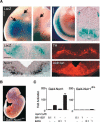
Figure 1.
Figure 1.
Gal4-Nurr1- and RXR-dependent activity localized in the ventral midbrain of Gal4-Nurr1 transgenic mice. (A, Top panel) Closeups of whole-mount X-gal-stained Gal4-Nurr1 embryos displaying blue staining in the forebrain (lower arrow, top left picture), VMB (upper arrow, top left picture), medulla (middle left), and spinal cord (middle right). Cross-section through the spinal cord revealed the main staining to be localized ventrally and dorsolaterally (right picture). Bars: left, 480 μm (also applies for middle left); middle right, 1000 μm; and right, 140 μm. (Middle and lower panels) Coronal sections from a representative Gal4-Nurr1 transgenic embryo at E11.5 showing X-gal staining in the ventral midbrain (left, top section) in the region where also the dopaminergic markers Nurr1 mRNA (left, bottom section) and TH immunoreactivity (right, top section) can be seen, as well as retinaldehydrogenase Aldh1a1 (right, bottom section), which is expressed in the DA progenitor cells. Bar, 100 μm. (B) Whole-mount, representative picture of Gal4-Nurr1dim embryo, which lacks blue X-gal staining in the entire CNS. Bar, 1500 μm. (C) Treatment of transiently transfected JEG3 cells. Gal4-Nurr1 or Gal4-Nurr1dim and human RXRα expression vectors were cotransfected with a luciferase reporter gene containing Gal4-binding sites. An expression vector expressing β-galactosidase was used as an internal control. Cells were treated with or without RXR ligands.
Figure 2.
Figure 2.
Wild-type ventral midbrain tissue activates a reporter gene assay in an RXR-dependent manner. (A,B) Excised tissue from E12.5 dorsal and ventral midbrain of wild-type mice was placed on JEG-3 cells transiently transfected as in Fig. 1 with Gal4-Nurr1 and Gal4-Nurr1dim expression vectors, respectively. Activity of the reporter gene was analyzed 32 h posttransfection. Actual luciferase values vary between experiments because of differences in transfection efficiencies. Values are therefore given as “Fold activation.” Results were consistent in at least three independent experiments. The value 1 is set as the activation obtained with indicated Gal4 constructs cotransfected with luciferase reporter but without addition of ligands or conditioned medium. (A) Ventral midbrain tissue activates Gal4-Nurr1, whereas dorsal midbrain tissue did not result in significant reporter gene activation. Typically, E13.5 ventral midbrain tissue activated at 10%-30% of the level achieved with 1 μM SR11237 (129-fold activation with SR11237 in the displayed experiment). (B) Addition of the RXR-specific antagonist LG849 blocked activation by Gal4-Nurr1 in cells cultured together with ventral midbrain tissue. (C) Reporter gene analysis of VMB activity from E13.5, E14.5, and E15.5 embryos shows that it increases with age. Very high levels of activation are seen when E15.5-conditioned medium is administered to transfected cells. In this particular experiment, E15.5-ventral-tissue-conditioned medium resulted in activation at levels seen with 1 μM SR11237. (D) Midbrain and cortex tissues do not activate RAR. Activity of midbrain and cortex tissue was assayed as above in cells expressing either Gal4-RAR or Gal4-Nurr1, respectively. (E) Partial purification of tissue-derived activity from mouse midbrain/cortex and analysis by negative ion mass spectrometry. Hexane extract as well as the most active HPLC fraction contained activity that stimulated cotransfected Gal4-Nurr1/RXR but not Gal4-RAR. (F) DHA was the main constituent in the most active fraction (#4). The arrow indicates the predicted mass for 9cis RA. As seen from the diagram, no detectable levels of 9cis RA were identified in this fraction.
Figure 3.
Figure 3.
RXR-specific ligands increase the number of surviving DA neurons. Primary cultures from rat ventral midbrain were established at E14.5-E15.5, incubated for 3-5 d in the presence of various ligands, fixed, and stained for DA neuron marker expression. Incubation in N2 media without any addition of ligand was used as control. (A) A few percent of the ventral midbrain cells are DA neurons according to cytoplasmic expression of TH (brightfield; left panel). The TH-positive cells express Nurr1 in the nucleus and Aldh1a1 in the cytoplasm. (B) Treatment with LG268 at 0.001-0.3 μM increases the number of TH-positive cells in a dose-dependent manner. Similar treatments with SR11237 also result in an increase in DA neuron number. (C) The effect of SR11237 and LG268 (both at 0.1 μM) was compared with that of GDNF (30 ng/mL). (D) Cultures were treated with LG268 alone and in combination with LG1208, an RXR-selective antagonist. Addition of LG1208 blocked the effect of LG268. Treatment of the cultures with LG1208 alone did not have a significant effect on the number of surviving cells. (**) p < 0.01; (***) p < 0.001. (E) To verify the activation potential of LG268 in neuronal cells, primary cultures were transfected with Gal4-Nurr1 and a Gal4-responsive luciferase vector. Values are given as “Fold activation,” where 1 is set as the activation obtained with Gal4-Nurr1 (or Gal4-Nurr1dim) cotransfected with the luciferase reporter but without addition of ligands. Gal4-Nurr1 is activated by LG268, whereas the ligand is unable to activate Gal4-Nurr1dim in neuronal cells. The RXR antagonist LG1208 completely abolished activation by LG268.
Figure 4.
Figure 4.
All-trans RA and the low-affinity RXR ligand DHA have opposite effects on DA neuron survival. (A) Treatment of the ventral midbrain primary cultures with 9cis RA did not result in an increase of the number of TH-positive cells (as shown with 10 and 30 nM). Addition of an RAR-selective antagonist Ro-415253 (RO; 10 μM) together with 9cis RA leads to a 100% increase in TH-positive cells. Treatment with RO alone did not increase the number of DA neurons. (B) Treatment of the primary cultures with the RAR-specific agonist TTNPB did not increase DA cell survival (at 10, 30, and 100 nM). Addition of TTNPB together with LG268 blocked the survival effect seen by addition of LG268 alone. (C,D) Primary midbrain cultures were treated with DHA (10, 30, and 100 μM). A dose-dependent increase in the number of TH-positive cells was observed. Addition of the RXR antagonist LG1208 (10 μM) together with DHA (10 and 100 μM) blocked the effect on cell survival. (*) p < 0.05; (**) p < 0.01; (***) p < 0.001.
Figure 5.
Figure 5.
RXR ligands do not cause a general increase in neuronal survival. (A,B) VMB cultures were treated with 0.1 μM LG268 or 60 μM DHA, left for 3 d, and then stained for NeuN IR to detect the total neuronal population. LG268 and DHA did not influence the survival of the general neuronal population as no significant changes in the number of neurons were seen by the RXR ligand treatments.
Figure 6.
Figure 6.
The RXR-dependent survival effect is mediated via Nurr1. E15.5 rat cortex, and hippocampus primary cultures were incubated for 3 d with or without 0.1 μM LG268, fixed, and stained for Nurr1 expression. (A) Brightfield images of cortical (left) and hippocampal (right) cultures analyzed for Nurr1 IR. (B) Approximately one-third of the cells are Nurr1 IR in the cortical cultures. In response to treatment with 0.1 μM LG268, the number of Nurr1 IR cells increased (left panel), whereas the number of non-Nurr1 IR cells did not (right panel). (C) A similar effect was observed in hippocampal cultures. (D) Cortical cultures were prepared from Nurr1+/+ and Nurr1-/- E15.5 embryos and incubated with or without 0.1 μM LG268. In cultures from Nurr1+/+ embryos, the total neuronal population as determined by NeuN expression increased in response to LG268 treatment, but no such increase was observed in cultures from Nurr1-/- embryos. (**) p < 0.01; (***) p < 0.001; (CTX) cortex; (HC) hippocampus.
Figure 7.
Figure 7.
A Nurr1/RXR-selective ligand increases cell survival. (A) In CV-1 cells, several Gal4-NR constructs were cotransfected with RXR. XCT0135908 (1 μM; the structure is shown in A) selectively increased the luciferase activity ∼37-fold when added to cells transfected with the Gal4-Nurr1 and a full-length RXRα expression vectors. Addition of this ligand did not increase the activity in cells transfected with any of the other Gal4-NRs, including Gal4-RXR, showing that XCT0135908 is strictly selective for RXR/Nurr1 heterodimers at 1 μM. (B) Addition of 1 μM XCT0135908 to rat VMB primary cultures increased the number of surviving TH-positive cells by 45% compared with N2 media. (***) p < 0.001. (C) To verify the activation potential of XCT0135908 in neuronal cells, primary cultures were transfected with Gal4-Nurr1 and a Gal4-responsive luciferase vector. Values are given as “Fold activation,” where 1 is set as the activation obtained with Gal4-Nurr1 (or Gal4-Nurr1dim) cotransfected with luciferase reporter but without addition of ligands. Gal4-Nurr1 is activated by XCT0135908, whereas the ligand is unable to activate Gal4-Nurr1dim in neuronal cells. The RXR antagonist LG1208 completely abolished activation by XCT0135908.

References

    1. Aarnisalo P., Kim, C.H., Lee, J.W., and Perlmann, T. 2002. Defining requirements for heterodimerization between the retinoid X receptor and the orphan nuclear receptor Nurr1. J. Biol. Chem. 277: 35118-35123. - PubMed
    1. Apfel C., Bauer, M., Crettaz, M., Forni, L., Kamber, M., Kaufman, F., LeMotte, P., Pirson, W., and Klaus, M. 1992. A retinoic acid receptor α antagonist selectively counteracts retinoic acid effects. Proc. Natl. Acad. Sci. 89: 7129-7133. - PMC - PubMed
    1. Aranda A. and Pascual, A. 2001. Nuclear hormone receptors and gene expression. Physiol. Rev. 81: 1269-1304. - PubMed
    1. Baker R.R. and Chang, H.Y. 1992. The hydrolysis of natural phosphatidylethanolamines by phospholipase A2 from rat serum: A degree of selectivity is shown for docosahexaenoate release. Biochim. Biophys. Acta 1125: 56-61. - PubMed
    1. Baker K.D., Shewchuk, L.M., Kozlova, T., Makishima, M., Hassell, A., Wisely, B., Caravella, J.A., Lambert, M.H., Reinking, J.L., Krause, H., et al. 2003. The Drosophila orphan nuclear receptor DHR38 mediates an atypical ecdysteroid signaling pathway. Cell 113: 731-742. - PubMed

Publication types

MeSH terms

LinkOut - more resources