. 2004 Jan 1;32(Database issue):D497-501.
doi: 10.1093/nar/gkh070.

Human protein reference database as a discovery resource for proteomics

Affiliations

Human protein reference database as a discovery resource for proteomics

Suraj Peri et al. Nucleic Acids Res. .

Abstract

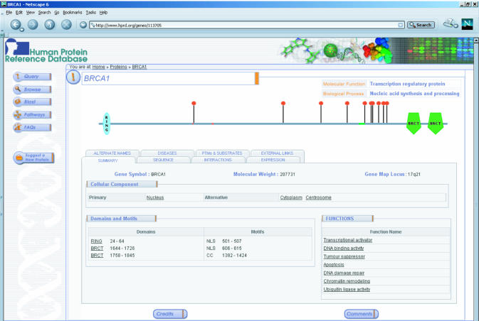
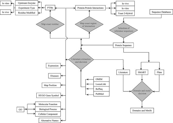
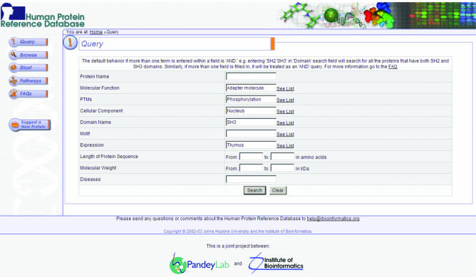
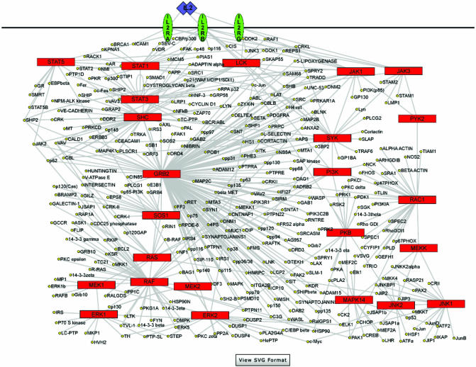
The rapid pace at which genomic and proteomic data is being generated necessitates the development of tools and resources for managing data that allow integration of information from disparate sources. The Human Protein Reference Database (http://www.hprd.org) is a web-based resource based on open source technologies for protein information about several aspects of human proteins including protein-protein interactions, post-translational modifications, enzyme-substrate relationships and disease associations. This information was derived manually by a critical reading of the published literature by expert biologists and through bioinformatics analyses of the protein sequence. This database will assist in biomedical discoveries by serving as a resource of genomic and proteomic information and providing an integrated view of sequence, structure, function and protein networks in health and disease.


Figures

Figure 1
Figure 1
A screenshot of the molecule page for BRCA1. The molecule page serves as the entry point for accessing detailed annotation of proteins in HPRD. Molecular function and biological process for each molecule are indicated at the top right with a graphic representation of the protein domains and motifs and PTMs displayed in the middle. Under the summary tab, the HUGO gene symbol, the calculated molecular weight and the gene locus are indicated along with the cellular component, which refers to subcellular localization. The extent of domains and motifs is shown under the summary tab but can also be accessed by mousing over an individual region or motif in the graph. All underlined text is linked to PubMed entries that provide further details or to a database source.
Figure 2
Figure 2
A schematic outlining the basic steps in the annotation procedure. The annotations are carried out by trained biologists who critically read the published literature. The entries to be annotated are carefully selected from all the existing database entries by BLAST analysis (6) as well as manual inspection to provide the reference sequence. Interpretive annotation steps are represented by orange diamonds in the schematic. For instance, the annotator performs domain and motif analysis using the SMART (7) and Pfam (8) programs as well as by reading the literature. The OMIM database is used for disease annotations (9), and RefSeq and LocusLink (10) for sequences and links to other databases.
Figure 3
Figure 3
A screenshot of the query page in HPRD. A user can search multiple annotation fields to retrieve protein entries based on protein name, molecular function, PTMs, cellular component, domains, motifs, tissue expression, length, molecular weight and diseases. The query system allows Boolean searches and the query terms are provided as pop-up lists for query fields other than protein name and diseases. Wild card searches using * are permitted by the query engine. Querying multiple search fields is processed as an ‘AND’ type of query by default. For example, a search for an adapter molecule, phosphorylation, nucleus, SH3 domain and thymus, as shown in the figure will retrieve a single entry for the Fyn binding protein, FYB, which satisfies all these parameters.
Figure 4
Figure 4
Visualizing protein interaction networks in HPRD. The interleukin-2 receptor pathway is shown with red boxes indicating the major signaling molecules in the pathway and yellow circles representing the interaction partners. The pathways are displayed as jpeg images by default with the option of viewing them as SVG images by clicking on the ‘view SVG format’ button at the bottom. With an appropriate SVG viewer plug-in, a user is able to visualize the network with additional functionalities such as zoom operations, a search function within the pathway, a hand tool for maneuvering the figure and links to individual molecule pages by clicking on the name of any protein.

References

    1. The International Human Genome Sequencing Consortium (2001) Initial sequencing and analysis of the human genome. Nature, 409, 860–921. - PubMed
    1. Venter J.C., Adams,M.D., Myers,E.W., Li,P.W., Mural,R.J., Sutton,G.G., Smith,H.O., Yandell,M., Evans,C.A., Holt,R.A. et al. (2001) The sequence of the human genome. Science, 291, 1304–1351. - PubMed
    1. Mann M. and Pandey,A. (2001) Use of mass spectrometry-derived data to annotate nucleotide and protein sequence databases. Trends Biochem. Sci., 26, 54–61. - PubMed
    1. Tucker C.L., Gera,J.F. and Uetz,P. (2001) Towards an understanding of complex protein networks. Trends Cell Biol., 11, 102–116. - PubMed
    1. Peri S., Navarro,J.D., Amanchy,R., Kristiansen,T.Z., Jonnalagadda,C.K., Surendranath,V., Niranjan,V., Muthusamy,B., Gandhi,T.K.B., Gronborg,M. et.al. (2003) Development of human protein reference database as an initial platform for approaching systems biology in humans. Genome Res., 13, 2363–2371. - PMC - PubMed