. 2003 Dec;54(6):769-80.
doi: 10.1002/ana.10762.

Insertion of mutant proteolipid protein results in missorting of myelin proteins

Affiliations

Insertion of mutant proteolipid protein results in missorting of myelin proteins

Catherine Vaurs-Barriere et al. Ann Neurol. 2003 Dec.

Erratum in

  • Ann Neurol. 2004 Jan;55(1):149-50

Abstract

Two brothers with a leukodystrophy, progressive spastic diplegia, and peripheral neuropathy were found to have proteinaceous aggregates in the peripheral nerve myelin sheath. The patients' mother had only subclinical peripheral neuropathy, but the maternal grandmother had adult-onset leukodystrophy. Sequencing of the proteolipid protein (PLP) gene showed a point mutation IVS4 + 1 G-->A within the donor splice site of intron 4. We identified one transcript with a deletion of exon 4 (Deltaex4, 169bp) encoding for PLP and DM20 proteins and lacking two transmembrane domains, and a second transcript with exon 4 + 10bp encoding three transmembrane domains. Immunohistochemistry showed abnormal aggregation in the myelin sheath of MBP and P0. Myelin-associated glycoprotein was present in the Schmidt-Lanterman clefts but significantly reduced in the periaxonal region. Using immunogold electron microscopy, we demonstrated the presence of mutated PLP/DM20 and the absence of the intact protein in the patient peripheral myelin sheath. We conclude that insertion of mutant PLP/DM20 with resulting aberrant distribution of other myelin proteins in peripheral nerve may constitute an important mechanism of dysmyelination in disorders associated with PLP mutations.


Figures

Fig 1
Fig 1
The pedigree of the affected family. The carrier status of Patient III-1 is not known.
Fig 2
Fig 2
Aberrant splicing resulting from the mutation IVS4 + 1 G → A. (A) Reverse transcription polymerase chain reaction (PCR) products from total RNA extracted from sural nerve biopsy of patient and control with F1/R1 primers. Both proteolipid protein (PLP) and DM20 (missing 105bp of exon 3b) transcripts were found in the control, with DM20 more abundant than the full-length PLP transcript. In the patient sample, three different transcripts were found, one corresponding in size to wild-type DM20, and two shorter products. Sequencing showed that the two smaller products correspond to PLP and DM20 with deletion of exon 4 (169bp), whereas the larger one corresponded to DM20 containing exon 4 plus 10bp from intron 4. (B) Ex3bF/Ex4R PCR amplification on F1/R1 PCR product from patient shows that a PLP transcript containing exon 4 is also expressed. (C) Schematic overview showing the position of IVS4 + 1 G → A mutation in the patient and the different resulting splice forms. Donor and acceptor splice sites are indicated in capital letters. The underlined GT sequence in intron 4 corresponds to the new splice donor site used to splice exon 4 + 10bp with exon 5. (D) Two types of mutated proteins were expressed in the patient; one containing 178 amino acids of PLP (or 143 amino acids of DM20) with an additional 27 frameshifted amino acid sequence (marked in black) at the C terminus, and the other containing 208 amino acids of PLP (or 173 amino acids of DM20) with an additional 26 frame-shifted amino acid sequence at the C terminus.
Fig 2
Fig 2
Aberrant splicing resulting from the mutation IVS4 + 1 G → A. (A) Reverse transcription polymerase chain reaction (PCR) products from total RNA extracted from sural nerve biopsy of patient and control with F1/R1 primers. Both proteolipid protein (PLP) and DM20 (missing 105bp of exon 3b) transcripts were found in the control, with DM20 more abundant than the full-length PLP transcript. In the patient sample, three different transcripts were found, one corresponding in size to wild-type DM20, and two shorter products. Sequencing showed that the two smaller products correspond to PLP and DM20 with deletion of exon 4 (169bp), whereas the larger one corresponded to DM20 containing exon 4 plus 10bp from intron 4. (B) Ex3bF/Ex4R PCR amplification on F1/R1 PCR product from patient shows that a PLP transcript containing exon 4 is also expressed. (C) Schematic overview showing the position of IVS4 + 1 G → A mutation in the patient and the different resulting splice forms. Donor and acceptor splice sites are indicated in capital letters. The underlined GT sequence in intron 4 corresponds to the new splice donor site used to splice exon 4 + 10bp with exon 5. (D) Two types of mutated proteins were expressed in the patient; one containing 178 amino acids of PLP (or 143 amino acids of DM20) with an additional 27 frameshifted amino acid sequence (marked in black) at the C terminus, and the other containing 208 amino acids of PLP (or 173 amino acids of DM20) with an additional 26 frame-shifted amino acid sequence at the C terminus.
Fig 3
Fig 3
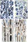
Light microscopy, ultrastructure, and immunohistochemistry of biopsied sural nerve. Light microscopy of toluidine blue–stained 1µm sections of control (A) with the normal thickness of myelin sheaths (arrow) and the patient (B), showing many thinly myelinated axons (arrow), loss of compaction of myelin sheaths and possible intramyelinic particulate material (B, arrowhead). (C) Immunohistochemical staining for P0 (C, control; D, patient) and myelin basic protein (E, control; F, patient) showing uneven and aggregated/particulate distribution (arrows) of these myelin proteins in the patient compared with the uniform staining in the control (arrows). (G) control and (H) patient nerves stained for myelin-associated glycoprotein showing normal continuous periaxonal (arrows) and Schmidt–Lanterman cleft staining (arrowheads) in the control. Myelin-associated glycoprotein reactivity is absent from the periaxonal region (arrows) and is present only in the Schmidt–Lanterman clefts (arrowheads) in the patient’s nerve. (I) Western blot of myelin proteins in patient (Pt) and control (Con) showing a marked reduction of these proteins in the patient. Proteolipid protein and DM20 were undetectable using this method (data not shown). Original magnification ×200 in A and B; ×400 in C to F; and ×1,000 oil immersion lens in G and H.
Fig 3
Fig 3
Light microscopy, ultrastructure, and immunohistochemistry of biopsied sural nerve. Light microscopy of toluidine blue–stained 1µm sections of control (A) with the normal thickness of myelin sheaths (arrow) and the patient (B), showing many thinly myelinated axons (arrow), loss of compaction of myelin sheaths and possible intramyelinic particulate material (B, arrowhead). (C) Immunohistochemical staining for P0 (C, control; D, patient) and myelin basic protein (E, control; F, patient) showing uneven and aggregated/particulate distribution (arrows) of these myelin proteins in the patient compared with the uniform staining in the control (arrows). (G) control and (H) patient nerves stained for myelin-associated glycoprotein showing normal continuous periaxonal (arrows) and Schmidt–Lanterman cleft staining (arrowheads) in the control. Myelin-associated glycoprotein reactivity is absent from the periaxonal region (arrows) and is present only in the Schmidt–Lanterman clefts (arrowheads) in the patient’s nerve. (I) Western blot of myelin proteins in patient (Pt) and control (Con) showing a marked reduction of these proteins in the patient. Proteolipid protein and DM20 were undetectable using this method (data not shown). Original magnification ×200 in A and B; ×400 in C to F; and ×1,000 oil immersion lens in G and H.
Fig 4
Fig 4
Immunoelectron microscopy. Immunoelectron microscopy for proteolipid protein (PLP) protein of nerve biopsy tissue of control (A) and patient (B) using secondary antibodies tagged with either 5nm gold particles for PLP-specific residues 117 to 129 (small arrow in both main image and inset) and 10nm gold particles recognizing residues 40 to 59 of both PLP and DM20 (large arrow in both main image and inset). (B) The mutant protein is identified both in the normal and in the poorly compacted myelin.

References

    1. Yool DA, Edgar JM, Montague P, Malcolm S. The proteolipid protein gene and myelin disorders in man and animal models. Hum Mol Genet. 2000;9:987–992. - PubMed
    1. Sporkel O, Uschkureit T, Bussow H, Stoffel W. Oligodendrocytes expressing exclusively the DM20 isoform of the proteolipid protein gene: myelination and development. Glia. 2002;37:19–30. - PubMed
    1. Klugmann M, Schwab MH, Puhlhofer A, et al. Assembly of CNS myelin in the absence of proteolipid protein. Neuron. 1997;18:59–70. - PubMed
    1. Campagnoni AT, Skoff RP. The pathobiology of myelin mutants reveal novel biological functions of the MBP and PLP genes. Brain Pathol. 2001;11:74–91. - PMC - PubMed
    1. Cailloux F, Gauthier-Barichard F, Mimault C, et al. Genotype-phenotype correlation in inherited brain myelination defects due to proteolipid protein gene mutations. Clin Eur Network Brain Dysmyelinating Dis Eur J Hum Genet. 2000;8:837–845. - PubMed

Publication types